| molecular function |
|---|
| | GO:0016787 | | hydrolase activity | | Catalysis of the hydrolysis of various bonds, e.g. C-O, C-N, C-C, phosphoric anhydride bonds, etc. Hydrolase is the systematic name for any enzyme of EC class 3. |
| | GO:0016791 | | phosphatase activity | | Catalysis of the hydrolysis of phosphoric monoesters, releasing inorganic phosphate. |
| | GO:0004721 | | phosphoprotein phosphatase activity | | Catalysis of the reaction: a phosphoprotein + H2O = a protein + phosphate. Together with protein kinases, these enzymes control the state of phosphorylation of cell proteins and thereby provide an important mechanism for regulating cellular activity. |
| | GO:0019901 | | protein kinase binding | | Interacting selectively and non-covalently with a protein kinase, any enzyme that catalyzes the transfer of a phosphate group, usually from ATP, to a protein substrate. |
| | GO:0004725 | | protein tyrosine phosphatase activity | | Catalysis of the reaction: protein tyrosine phosphate + H2O = protein tyrosine + phosphate. |
| biological process |
|---|
| | GO:0030030 | | cell projection organization | | A process that is carried out at the cellular level which results in the assembly, arrangement of constituent parts, or disassembly of a prolongation or process extending from a cell, e.g. a flagellum or axon. |
| | GO:0060271 | | cilium assembly | | The assembly of a cilium, a specialized eukaryotic organelle that consists of a filiform extrusion of the cell surface. Each cilium is bounded by an extrusion of the cytoplasmic membrane, and contains a regular longitudinal array of microtubules, anchored basally in a centriole. |
| | GO:0016311 | | dephosphorylation | | The process of removing one or more phosphoric (ester or anhydride) residues from a molecule. |
| | GO:0010633 | | negative regulation of epithelial cell migration | | Any process that stops, prevents, or reduces the frequency, rate or extent of epithelial cell migration. |
| | GO:0035335 | | peptidyl-tyrosine dephosphorylation | | The removal of phosphoric residues from peptidyl-O-phospho-tyrosine to form peptidyl-tyrosine. |
| | GO:1903393 | | positive regulation of adherens junction organization | | Any process that activates or increases the frequency, rate or extent of adherens junction organization. |
| | GO:2000643 | | positive regulation of early endosome to late endosome transport | | Any process that activates or increases the frequency, rate or extent of early endosome to late endosome transport. |
| | GO:1903387 | | positive regulation of homophilic cell adhesion | | Any process that activates or increases the frequency, rate or extent of homophilic cell adhesion. |
| | GO:0006470 | | protein dephosphorylation | | The process of removing one or more phosphoric residues from a protein. |
| | GO:0015031 | | protein transport | | The directed movement of proteins into, out of or within a cell, or between cells, by means of some agent such as a transporter or pore. |
| | GO:0030334 | | regulation of cell migration | | Any process that modulates the frequency, rate or extent of cell migration. |
| | GO:0006810 | | transport | | The directed movement of substances (such as macromolecules, small molecules, ions) or cellular components (such as complexes and organelles) into, out of or within a cell, or between cells, or within a multicellular organism by means of some agent such as a transporter, pore or motor protein. |
| | GO:0043162 | | ubiquitin-dependent protein catabolic process via the multivesicular body sorting pathway | | The chemical reactions and pathways resulting in the breakdown of a protein or peptide covalently tagged with ubiquitin, via the multivesicular body (MVB) sorting pathway; ubiquitin-tagged proteins are sorted into MVBs, and delivered to a lysosome/vacuole for degradation. |
| cellular component |
|---|
| | GO:0042995 | | cell projection | | A prolongation or process extending from a cell, e.g. a flagellum or axon. |
| | GO:0036064 | | ciliary basal body | | A membrane-tethered, short cylindrical array of microtubules and associated proteins found at the base of a eukaryotic cilium (also called flagellum) that is similar in structure to a centriole and derives from it. The cilium basal body is the site of assembly and remodelling of the cilium and serves as a nucleation site for axoneme growth. As well as anchoring the cilium, it is thought to provide a selective gateway regulating the entry of ciliary proteins and vesicles by intraflagellar transport. |
| | GO:0005929 | | cilium | | A specialized eukaryotic organelle that consists of a filiform extrusion of the cell surface and of some cytoplasmic parts. Each cilium is largely bounded by an extrusion of the cytoplasmic (plasma) membrane, and contains a regular longitudinal array of microtubules, anchored to a basal body. |
| | GO:0005737 | | cytoplasm | | All of the contents of a cell excluding the plasma membrane and nucleus, but including other subcellular structures. |
| | GO:0031410 | | cytoplasmic vesicle | | A vesicle found in the cytoplasm of a cell. |
| | GO:0005856 | | cytoskeleton | | Any of the various filamentous elements that form the internal framework of cells, and typically remain after treatment of the cells with mild detergent to remove membrane constituents and soluble components of the cytoplasm. The term embraces intermediate filaments, microfilaments, microtubules, the microtrabecular lattice, and other structures characterized by a polymeric filamentous nature and long-range order within the cell. The various elements of the cytoskeleton not only serve in the maintenance of cellular shape but also have roles in other cellular functions, including cellular movement, cell division, endocytosis, and movement of organelles. |
| | GO:0005769 | | early endosome | | A membrane-bounded organelle that receives incoming material from primary endocytic vesicles that have been generated by clathrin-dependent and clathrin-independent endocytosis; vesicles fuse with the early endosome to deliver cargo for sorting into recycling or degradation pathways. |
| | GO:0005768 | | endosome | | A vacuole to which materials ingested by endocytosis are delivered. |
| | GO:0070062 | | extracellular exosome | | A vesicle that is released into the extracellular region by fusion of the limiting endosomal membrane of a multivesicular body with the plasma membrane. Extracellular exosomes, also simply called exosomes, have a diameter of about 40-100 nm. |
| | GO:0005654 | | nucleoplasm | | That part of the nuclear content other than the chromosomes or the nucleolus. |
| | GO:0005634 | | nucleus | | A membrane-bounded organelle of eukaryotic cells in which chromosomes are housed and replicated. In most cells, the nucleus contains all of the cell's chromosomes except the organellar chromosomes, and is the site of RNA synthesis and processing. In some species, or in specialized cell types, RNA metabolism or DNA replication may be absent. |
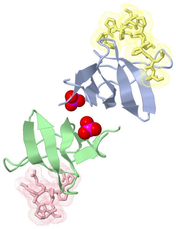
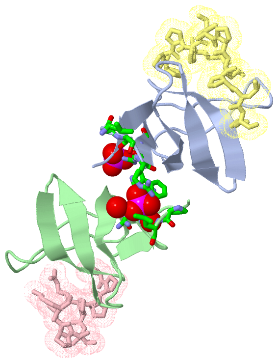
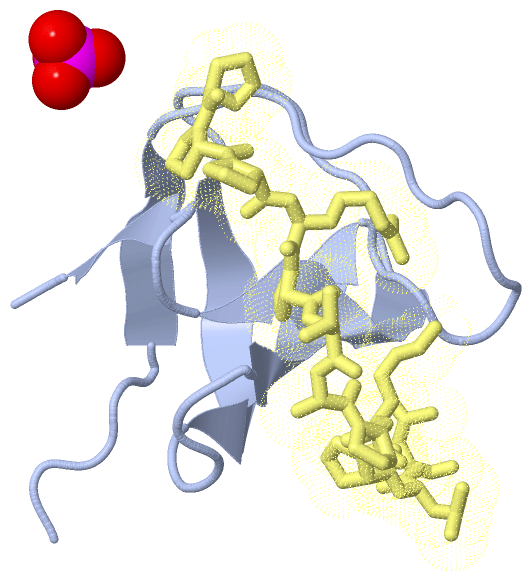
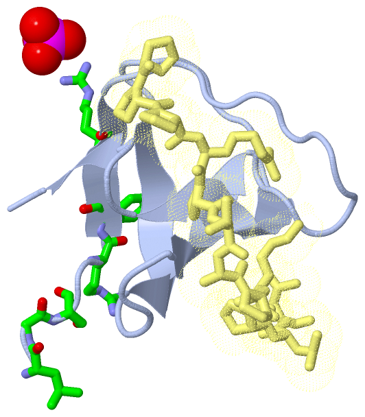
 Description