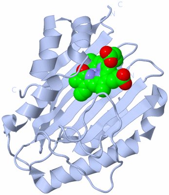

|
|
|

 Description
|
|

 Compounds
|
||||||||||||||||||||||||||||||||||||||||||||

 Chains, Units
Summary Information (see also Sequences/Alignments below) |

 Ligands, Modified Residues, Ions (1, 4)
Asymmetric Unit (1, 4)
|

 Sites (4, 4)
Asymmetric Unit (4, 4)
|

 SS Bonds (0, 0)
| (no "SS Bond" information available for 2VGR) |

 Cis Peptide Bonds (8, 8)
Asymmetric Unit
|
||||||||||||||||||||||||||||||||||||

 SAPs(SNPs)/Variants (0, 0)
| (no "SAP(SNP)/Variant" information available for 2VGR) |

 PROSITE Motifs (0, 0)
| (no "PROSITE Motif" information available for 2VGR) |

 Exons (0, 0)
| (no "Exon" information available for 2VGR) |

 Sequences/Alignments
Asymmetric UnitChain A from PDB Type:PROTEIN Length:210 aligned with PEBS_BPPRM | Q58MU6 from UniProtKB/Swiss-Prot Length:233 Alignment length:213 30 40 50 60 70 80 90 100 110 120 130 140 150 160 170 180 190 200 210 220 230 PEBS_BPPRM 21 KTIWQNYIDALFETFPQLEISEVWAKWDGGNVTKDGGDAKLTANIRTGEHFLKAREAHIVDPNSDIYNTILYPKTGADLPCFGMDLMKFSDKKVIIVFDFQHPREKYLFSVDGLPEDDGKYRFFEMGNHFSKNIFVRYCKPDEVDQYLDTFKLYLTKYKEMIDNNKPVGEDTTVYSDFDTYMTELDPVRGYMKNKFGEGRSEAFVNDFLFSYK 233 SCOP domains --------------------------------------------------------------------------------------------------------------------------------------------------------------------------------------------------------------------- SCOP domains CATH domains --------------------------------------------------------------------------------------------------------------------------------------------------------------------------------------------------------------------- CATH domains Pfam domains --------------------------------------------------------------------------------------------------------------------------------------------------------------------------------------------------------------------- Pfam domains Sec.struct. author ...hhhhhhhhhhhh...eeeeeeeeeee...---...eeeeeeeeee..eeeeeeeeee....eeeeeeeee.......eeeeeeee.....eeeeeeee.................................eeeeehhhhhhhhhhhhhhhhhhhhhhhhhhh....hhhhhhhhhhhhh...hhhhhhhhhhhhhhhhhhhhhh..... Sec.struct. author SAPs(SNPs) --------------------------------------------------------------------------------------------------------------------------------------------------------------------------------------------------------------------- SAPs(SNPs) PROSITE --------------------------------------------------------------------------------------------------------------------------------------------------------------------------------------------------------------------- PROSITE Transcript --------------------------------------------------------------------------------------------------------------------------------------------------------------------------------------------------------------------- Transcript 2vgr A 21 KTIWQNYIDALFETFPQLEISEVWAKWDGGNV---GGDAKLTANIRTGEHFLKAREAHIVDPNSDIYNTILYPKTGADLPCFGMDLMKFSDKKVIIVFDFQHPREKYLFSVDGLPEDDGKYRFFEMGNHFSKNIFVRYCKPDEVDQYLDTFKLYLTKYKEMIDNNKPVGEDTTVYSDFDTYMTELDPVRGYMKNKFGEGRSEAFVNDFLFSYK 233 30 40 50 | | 60 70 80 90 100 110 120 130 140 150 160 170 180 190 200 210 220 230 52 56 Chain B from PDB Type:PROTEIN Length:211 aligned with PEBS_BPPRM | Q58MU6 from UniProtKB/Swiss-Prot Length:233 Alignment length:213 30 40 50 60 70 80 90 100 110 120 130 140 150 160 170 180 190 200 210 220 230 PEBS_BPPRM 21 KTIWQNYIDALFETFPQLEISEVWAKWDGGNVTKDGGDAKLTANIRTGEHFLKAREAHIVDPNSDIYNTILYPKTGADLPCFGMDLMKFSDKKVIIVFDFQHPREKYLFSVDGLPEDDGKYRFFEMGNHFSKNIFVRYCKPDEVDQYLDTFKLYLTKYKEMIDNNKPVGEDTTVYSDFDTYMTELDPVRGYMKNKFGEGRSEAFVNDFLFSYK 233 SCOP domains --------------------------------------------------------------------------------------------------------------------------------------------------------------------------------------------------------------------- SCOP domains CATH domains --------------------------------------------------------------------------------------------------------------------------------------------------------------------------------------------------------------------- CATH domains Pfam domains --------------------------------------------------------------------------------------------------------------------------------------------------------------------------------------------------------------------- Pfam domains Sec.struct. author ...hhhhhhhhhhhh...eeeeeeeeeee.....--..eeeeeeeeee..eeeeeeeeee....eeeeeeeee.......eeeeeeee.....eeeeeeee.................................eeeee...hhhhhhhhhhhhhhhhhhhhhhhh....hhhhhhhhhhhhhhh.hhhhhhhhhhhhhhhhhhhhhh..... Sec.struct. author SAPs(SNPs) --------------------------------------------------------------------------------------------------------------------------------------------------------------------------------------------------------------------- SAPs(SNPs) PROSITE --------------------------------------------------------------------------------------------------------------------------------------------------------------------------------------------------------------------- PROSITE Transcript --------------------------------------------------------------------------------------------------------------------------------------------------------------------------------------------------------------------- Transcript 2vgr B 21 KTIWQNYIDALFETFPQLEISEVWAKWDGGNVTK--GDAKLTANIRTGEHFLKAREAHIVDPNSDIYNTILYPKTGADLPCFGMDLMKFSDKKVIIVFDFQHPREKYLFSVDGLPEDDGKYRFFEMGNHFSKNIFVRYCKPDEVDQYLDTFKLYLTKYKEMIDNNKPVGEDTTVYSDFDTYMTELDPVRGYMKNKFGEGRSEAFVNDFLFSYK 233 30 40 50 | | 60 70 80 90 100 110 120 130 140 150 160 170 180 190 200 210 220 230 54 57 Chain C from PDB Type:PROTEIN Length:213 aligned with PEBS_BPPRM | Q58MU6 from UniProtKB/Swiss-Prot Length:233 Alignment length:215 28 38 48 58 68 78 88 98 108 118 128 138 148 158 168 178 188 198 208 218 228 PEBS_BPPRM 19 KSKTIWQNYIDALFETFPQLEISEVWAKWDGGNVTKDGGDAKLTANIRTGEHFLKAREAHIVDPNSDIYNTILYPKTGADLPCFGMDLMKFSDKKVIIVFDFQHPREKYLFSVDGLPEDDGKYRFFEMGNHFSKNIFVRYCKPDEVDQYLDTFKLYLTKYKEMIDNNKPVGEDTTVYSDFDTYMTELDPVRGYMKNKFGEGRSEAFVNDFLFSYK 233 SCOP domains ----------------------------------------------------------------------------------------------------------------------------------------------------------------------------------------------------------------------- SCOP domains CATH domains ----------------------------------------------------------------------------------------------------------------------------------------------------------------------------------------------------------------------- CATH domains Pfam domains ----------------------------------------------------------------------------------------------------------------------------------------------------------------------------------------------------------------------- Pfam domains Sec.struct. author .....hhhhhhhhhhhh...eeeeeeeeeeeeee.--.eeeeeeeeeeee..eeeeeeeeeee..eeeeeeeeee.......eeeeeeee.....eeeeeeee.................................eeeee...hhhhhhhhhhhhhhhhhhhhhhhh....hhhhhhhhhhhhhh..hhhhhhhhhhhhhhhhhhhhhh..... Sec.struct. author SAPs(SNPs) ----------------------------------------------------------------------------------------------------------------------------------------------------------------------------------------------------------------------- SAPs(SNPs) PROSITE ----------------------------------------------------------------------------------------------------------------------------------------------------------------------------------------------------------------------- PROSITE Transcript ----------------------------------------------------------------------------------------------------------------------------------------------------------------------------------------------------------------------- Transcript 2vgr C 19 KSKTIWQNYIDALFETFPQLEISEVWAKWDGGNVT--GGDAKLTANIRTGEHFLKAREAHIVDPNSDIYNTILYPKTGADLPCFGMDLMKFSDKKVIIVFDFQHPREKYLFSVDGLPEDDGKYRFFEMGNHFSKNIFVRYCKPDEVDQYLDTFKLYLTKYKEMIDNNKPVGEDTTVYSDFDTYMTELDPVRGYMKNKFGEGRSEAFVNDFLFSYK 233 28 38 48 | |58 68 78 88 98 108 118 128 138 148 158 168 178 188 198 208 218 228 53 56 Chain D from PDB Type:PROTEIN Length:213 aligned with PEBS_BPPRM | Q58MU6 from UniProtKB/Swiss-Prot Length:233 Alignment length:213 30 40 50 60 70 80 90 100 110 120 130 140 150 160 170 180 190 200 210 220 230 PEBS_BPPRM 21 KTIWQNYIDALFETFPQLEISEVWAKWDGGNVTKDGGDAKLTANIRTGEHFLKAREAHIVDPNSDIYNTILYPKTGADLPCFGMDLMKFSDKKVIIVFDFQHPREKYLFSVDGLPEDDGKYRFFEMGNHFSKNIFVRYCKPDEVDQYLDTFKLYLTKYKEMIDNNKPVGEDTTVYSDFDTYMTELDPVRGYMKNKFGEGRSEAFVNDFLFSYK 233 SCOP domains --------------------------------------------------------------------------------------------------------------------------------------------------------------------------------------------------------------------- SCOP domains CATH domains --------------------------------------------------------------------------------------------------------------------------------------------------------------------------------------------------------------------- CATH domains Pfam domains (1) ----Fe_bilin_red-2vgrD01 D:25-231 -- Pfam domains (1) Pfam domains (2) ----Fe_bilin_red-2vgrD02 D:25-231 -- Pfam domains (2) Pfam domains (3) ----Fe_bilin_red-2vgrD03 D:25-231 -- Pfam domains (3) Pfam domains (4) ----Fe_bilin_red-2vgrD04 D:25-231 -- Pfam domains (4) Sec.struct. author ...hhhhhhhhhhhh...eeeeeeeeeeeeee....eeeeeeeeeee...eeeeeeeeeee..eeeeeeeeee.......eeeeeeee.....eeeeeeee.................................eeeeehhhhhhhhhhhhhhhhhhhhhhhhhhh....hhhhhhhhhhhhhh....hhhhhhhhhhhhhhhhhhhh..... Sec.struct. author SAPs(SNPs) --------------------------------------------------------------------------------------------------------------------------------------------------------------------------------------------------------------------- SAPs(SNPs) PROSITE --------------------------------------------------------------------------------------------------------------------------------------------------------------------------------------------------------------------- PROSITE Transcript --------------------------------------------------------------------------------------------------------------------------------------------------------------------------------------------------------------------- Transcript 2vgr D 21 KTIWQNYIDALFETFPQLEISEVWAKWDGGNVTKDGGDAKLTANIRTGEHFLKAREAHIVDPNSDIYNTILYPKTGADLPCFGMDLMKFSDKKVIIVFDFQHPREKYLFSVDGLPEDDGKYRFFEMGNHFSKNIFVRYCKPDEVDQYLDTFKLYLTKYKEMIDNNKPVGEDTTVYSDFDTYMTELDPVRGYMKNKFGEGRSEAFVNDFLFSYK 233 30 40 50 60 70 80 90 100 110 120 130 140 150 160 170 180 190 200 210 220 230
|
||||||||||||||||||||

 SCOP Domains (0, 0)
| (no "SCOP Domain" information available for 2VGR) |

 CATH Domains (0, 0)
| (no "CATH Domain" information available for 2VGR) |

 Pfam Domains (1, 4)
Asymmetric Unit
 
|

 Gene Ontology (5, 5)
|
Asymmetric Unit(show GO term definitions) Chain A,B,C,D (PEBS_BPPRM | Q58MU6)
|
||||||||||||||||||||||||||||||||||||||||||

 Interactive Views
|
||||||||||||||||||||||||||||||||||||||||||||||||||||||||||||||||||||||||||||||||||||||||||||||||||||||||||||||||||||||||||||||||||||||||||||||||||||||||||||||||||||||||||||||||||||||||||||||||||||||||||||||||||||||||||||||

 Still Images
|
||||||||||||||||

 Databases
|
||||||||||||||||||||||||||||||||||||||||||||||||||||||||||||||||||||||||||||||||||||||||||||||||||||||||||||||||||||||||||||||||||||||||||||||||||||||||||||||||

 Analysis Tools
|
|||||||||||||||||||||||||||||||||||||||||||||||||||||||||||||

 Entries Sharing at Least One Protein Chain (UniProt ID)
 Related Entries Specified in the PDB File
|
|
|


