|
|
|
|
 Description
|
|
Compounds
|
||||||||||||||||||||||||||||||||||||||||||||
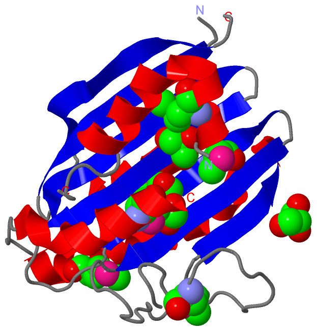
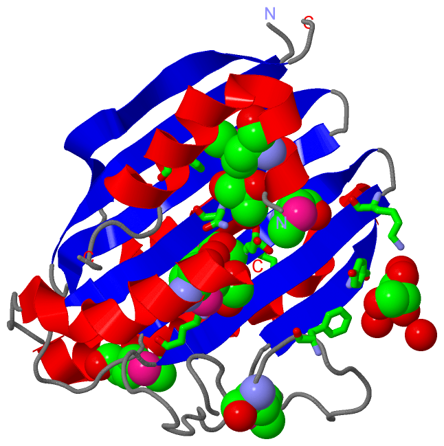
Chains, Units
Summary Information (see also Sequences/Alignments below) |
Ligands, Modified Residues, Ions (2, 9)| Asymmetric/Biological Unit (2, 9) |
Sites (3, 3)
Asymmetric Unit (3, 3)
|
SS Bonds (0, 0)| (no "SS Bond" information available for 2VCL) |
Cis Peptide Bonds (0, 0)| (no "Cis Peptide Bond" information available for 2VCL) |
SAPs(SNPs)/Variants (0, 0)| (no "SAP(SNP)/Variant" information available for 2VCL) |
PROSITE Motifs (0, 0)| (no "PROSITE Motif" information available for 2VCL) |
Exons (0, 0)| (no "Exon" information available for 2VCL) |
Sequences/Alignments
Asymmetric/Biological UnitChain A from PDB Type:PROTEIN Length:205 aligned with PEBS_BPPRM | Q58MU6 from UniProtKB/Swiss-Prot Length:233 Alignment length:212 31 41 51 61 71 81 91 101 111 121 131 141 151 161 171 181 191 201 211 221 231 PEBS_BPPRM 22 TIWQNYIDALFETFPQLEISEVWAKWDGGNVTKDGGDAKLTANIRTGEHFLKAREAHIVDPNSDIYNTILYPKTGADLPCFGMDLMKFSDKKVIIVFDFQHPREKYLFSVDGLPEDDGKYRFFEMGNHFSKNIFVRYCKPDEVDQYLDTFKLYLTKYKEMIDNNKPVGEDTTVYSDFDTYMTELDPVRGYMKNKFGEGRSEAFVNDFLFSYK 233 SCOP domains -------------------------------------------------------------------------------------------------------------------------------------------------------------------------------------------------------------------- SCOP domains CATH domains -------------------------------------------------------------------------------------------------------------------------------------------------------------------------------------------------------------------- CATH domains Pfam domains ---Fe_bilin_red-2vclA01 A:25-2 31 -- Pfam domains SAPs(SNPs) -------------------------------------------------------------------------------------------------------------------------------------------------------------------------------------------------------------------- SAPs(SNPs) PROSITE -------------------------------------------------------------------------------------------------------------------------------------------------------------------------------------------------------------------- PROSITE Transcript -------------------------------------------------------------------------------------------------------------------------------------------------------------------------------------------------------------------- Transcript 2vcl A 22 TIWQNYIDALFETFPQLEISEVWAKWDGGN-----GDAKLTANIRTGEHFLKAREAHIVDPNSDIYNTILYPKTGADLPCFGmDLmKFSDKKVIIVFDFQHPREKYLFSVDGLPEDDGKYRFFEmGNHFSKNIFVRYCKPDEVDQYLDTFKLYLTKYKEmIDNNKPVGEDTTVYSDFDTYmTEL--VRGYmKNKFGEGRSEAFVNDFLFSYK 233 31 41 51 | 61 71 81 91 101 | | 111 121 131 141 | 151 161 171 181 191 201| | |211| 221 231 51 57 104-MSE 146-MSE 181-MSE 202-MSE | | 107-MSE 205 | | 208 | 212-MSE
|
||||||||||||||||||||
SCOP Domains (0, 0)| (no "SCOP Domain" information available for 2VCL) |
CATH Domains (0, 0)| (no "CATH Domain" information available for 2VCL) |
Pfam Domains (1, 1)
Asymmetric/Biological Unit

|
Gene Ontology (5, 5)|
Asymmetric/Biological Unit(hide GO term definitions) Chain A (PEBS_BPPRM | Q58MU6)
|
||||||||||||||||||||||||||||||||||||||||||
Interactive Views
|
|||||||||||||||||||||||||||||||||||||||||||||||||||||||||||||||||||||||||||||||||||||||||||||||||||||||||||||||||||||||||||||||||||||||||||
Still Images
|
||||||||||||||||
Databases
|
||||||||||||||||||||||||||||||||||||||||||||||||||||||||||||||||||||||||||||||||||||||||||||||||||||||||||||||||||||||||||||||||||||||||||||||||||||||||||||||||
Analysis Tools
|
|||||||||||||||||||||||||||||||||||||||||||||||||||||||||||||
Entries Sharing at Least One Protein Chain (UniProt ID)
Related Entries Specified in the PDB File
|
|