|
|
|
|
 Description
|
|
Compounds
|
||||||||||||||||||||||||||||||||||||||||||||||||||||
Chains, Units
Summary Information (see also Sequences/Alignments below) |
Ligands, Modified Residues, Ions (5, 12)
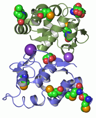
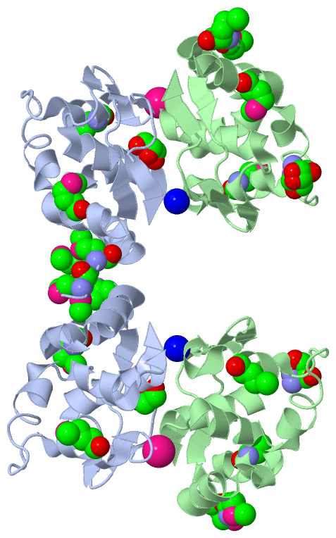
Asymmetric Unit (5, 12)
|
Sites (4, 4)
Asymmetric Unit (4, 4)
|
SS Bonds (0, 0)| (no "SS Bond" information available for 2QM2) |
Cis Peptide Bonds (0, 0)| (no "Cis Peptide Bond" information available for 2QM2) |
SAPs(SNPs)/Variants (0, 0)| (no "SAP(SNP)/Variant" information available for 2QM2) |
PROSITE Motifs (0, 0)| (no "PROSITE Motif" information available for 2QM2) |
Exons (0, 0)| (no "Exon" information available for 2QM2) |
Sequences/Alignments
Asymmetric UnitChain A from PDB Type:PROTEIN Length:118 aligned with Q87IM6_VIBPA | Q87IM6 from UniProtKB/TrEMBL Length:122 Alignment length:118 14 24 34 44 54 64 74 84 94 104 114 Q87IM6_VIBPA 5 FGMKMELQQFLDALASSPEKIEFETTMAVIEDNYDFTPAAFTNGNTQNDANENNGSCKIFAFGLLNALDKEATLACFGRFYREDVLLHPENNDHQNIRNFMVTGWEGIQFETSALTAK 122 SCOP domains ---------------------------------------------------------------------------------------------------------------------- SCOP domains CATH domains 2qm2A00 A:5-122 vpa0580 domain like CATH domains Pfam domains ---------------------------------------------------------------------------------------------------------------------- Pfam domains SAPs(SNPs) ---------------------------------------------------------------------------------------------------------------------- SAPs(SNPs) PROSITE ---------------------------------------------------------------------------------------------------------------------- PROSITE Transcript ---------------------------------------------------------------------------------------------------------------------- Transcript 2qm2 A 5 FGmKmELQQFLDALASSPEKIEFETTmAVIEDNYDFTPAAFTNGNTQNDANENNGSCKIFAFGLLNALDKEATLACFGRFYREDVLLHPENNDHQNIRNFmVTGWEGIQFETSALTAK 122 | | 14 24 | 34 44 54 64 74 84 94 104| 114 | | 31-MSE 105-MSE 7-MSE 9-MSE Chain B from PDB Type:PROTEIN Length:116 aligned with Q87IM6_VIBPA | Q87IM6 from UniProtKB/TrEMBL Length:122 Alignment length:116 16 26 36 46 56 66 76 86 96 106 116 Q87IM6_VIBPA 7 MKMELQQFLDALASSPEKIEFETTMAVIEDNYDFTPAAFTNGNTQNDANENNGSCKIFAFGLLNALDKEATLACFGRFYREDVLLHPENNDHQNIRNFMVTGWEGIQFETSALTAK 122 SCOP domains -------------------------------------------------------------------------------------------------------------------- SCOP domains CATH domains -2qm2B00 B:8-122 vpa0580 domain like CATH domains Pfam domains (1) --HopJ-2qm2B01 B:9-119 --- Pfam domains (1) Pfam domains (2) --HopJ-2qm2B02 B:9-119 --- Pfam domains (2) SAPs(SNPs) -------------------------------------------------------------------------------------------------------------------- SAPs(SNPs) PROSITE -------------------------------------------------------------------------------------------------------------------- PROSITE Transcript -------------------------------------------------------------------------------------------------------------------- Transcript 2qm2 B 7 mKmELQQFLDALASSPEKIEFETTmAVIEDNYDFTPAAFTNGNTQNDANENNGSCKIFAFGLLNALDKEATLACFGRFYREDVLLHPENNDHQNIRNFmVTGWEGIQFETSALTAK 122 | | 16 26 | 36 46 56 66 76 86 96 106 116 7-MSE 31-MSE 105-MSE 9-MSE
|
||||||||||||||||||||
SCOP Domains (0, 0)| (no "SCOP Domain" information available for 2QM2) |
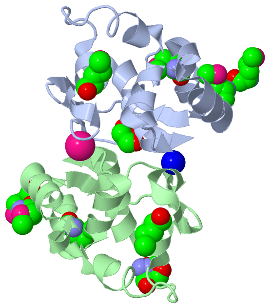
CATH Domains (1, 2)
Asymmetric Unit
 |
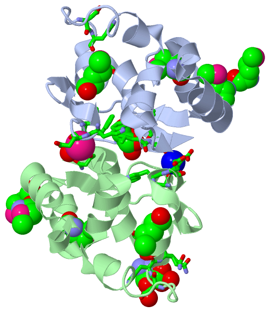
Pfam Domains (1, 2)
Asymmetric Unit
|
Gene Ontology (0, 0)|
Asymmetric Unit(hide GO term definitions)
(no "Gene Ontology" information available for 2QM2)
|
Interactive Views
|
||||||||||||||||||||||||||||||||||||||||||||||||||||||||||||||||||||||||||||||||||||||||||||||||||||||||||||||||||||||||||||||||||||||||||||||||||||||||||||||||||||||||||||||||||||||||||||||
Still Images
|
||||||||||||||||
Databases
|
||||||||||||||||||||||||||||||||||||||||||||||||||||||||||||||||||||||||||||||||||||||||||||||||||||||||||||||||||||||||||||||||||||||||||||||||||||||||||||||||
Analysis Tools
|
|||||||||||||||||||||||||||||||||||||||||||||||||||||||||||||
Entries Sharing at Least One Protein Chain (UniProt ID)
Related Entries Specified in the PDB File
|
|