Show PDB file:   
         Plain Text   HTML   (compressed file size)
QuickSearch:   
by PDB,NDB,UniProt,PROSITE Code or Search Term(s)  
(-)Asym./Biol. Unit
(-)Asym./Biol. Unit - sites
collapse expand < >
Image Asym./Biol. Unit
Asym./Biol. Unit  (Jmol Viewer)
Image Asym./Biol. Unit - sites
Asym./Biol. Unit - sites  (Jmol Viewer)

(-) Description

Title :  PVUII ENDONUCLEASE COMPLEXED TO AN IODINATED COGNATE DNA
 
Authors :  J. Horton, X. Cheng
Date :  01 Nov 98  (Deposition) - 20 Dec 99  (Release) - 15 Sep 09  (Revision)
Method :  X-RAY DIFFRACTION
Resolution :  1.76
Chains :  Asym./Biol. Unit :  A,B,C,D
Keywords :  Complex (Restriction Endonuclease/Dna), Hydrolase/Dna Complex (Keyword Search: [Gene Ontology, PubMed, Web (Google))
 
Reference :  J. R. Horton, J. Bonventre, X. Cheng
How Is Modification Of The Dna Substrate Recognized By The Pvuii Restriction Endonuclease?
J. Biol. Chem. V. 379 451 1998
PubMed-ID: 9628337
(for further references see the PDB file header)

(-) Compounds

Molecule 1 - DNA (5'-D(*TP*GP*AP*CP*CP*AP*GP*(C38) P*TP*GP*GP*TP*C)-3')
    ChainsC, D
    EngineeredYES
    SyntheticYES
 
Molecule 2 - TYPE II RESTRICTION ENZYME PVUII
    ChainsA, B
    EC Number3.1.21.4
    EngineeredYES
    Expression SystemESCHERICHIA COLI
    Expression System PlasmidPPR594
    Expression System StrainPR653
    Expression System Taxid562
    Organism ScientificPROTEUS VULGARIS
    Organism Taxid585
    SynonymENDONUCLEASE PVUII, R.PVUII

 Structural Features

(-) Chains, Units

  1234
Asymmetric/Biological Unit ABCD

Summary Information (see also Sequences/Alignments below)

(-) Ligands, Modified Residues, Ions  (1, 2)

Asymmetric/Biological Unit (1, 2)
No.NameCountTypeFull Name
1C382Mod. Nucleotide5-IODO-2'-DEOXY-CYTIDINE-5'-MONOPHOSPHATE

(-) Sites  (2, 2)

Asymmetric Unit (2, 2)
No.NameEvidenceResiduesDescription
1CAAAUTHORASP A:58 , GLU A:68 , LYS A:70CATALYTIC RESIDUES
2CABAUTHORASP B:58 , GLU B:68 , LYS B:70CATALYTIC RESIDUES

(-) SS Bonds  (0, 0)

(no "SS Bond" information available for 2PVI)

(-) Cis Peptide Bonds  (0, 0)

(no "Cis Peptide Bond" information available for 2PVI)

 Sequence-Structure Mapping

(-) SAPs(SNPs)/Variants  (0, 0)

(no "SAP(SNP)/Variant" information available for 2PVI)

(-) PROSITE Motifs  (0, 0)

(no "PROSITE Motif" information available for 2PVI)

(-) Exons   (0, 0)

(no "Exon" information available for 2PVI)

(-) Sequences/Alignments

Asymmetric/Biological Unit
   Reformat: Number of residues per line =  ('0' or empty: single-line sequence representation)
  Number of residues per labelling interval =   
  UniProt sequence: complete  aligned part    
   Show mapping: SCOP domains CATH domains Pfam domains Secondary structure (by author)
SAPs(SNPs) PROSITE motifs Exons
(details for a mapped element are shown in a popup box when the mouse pointer rests over it)
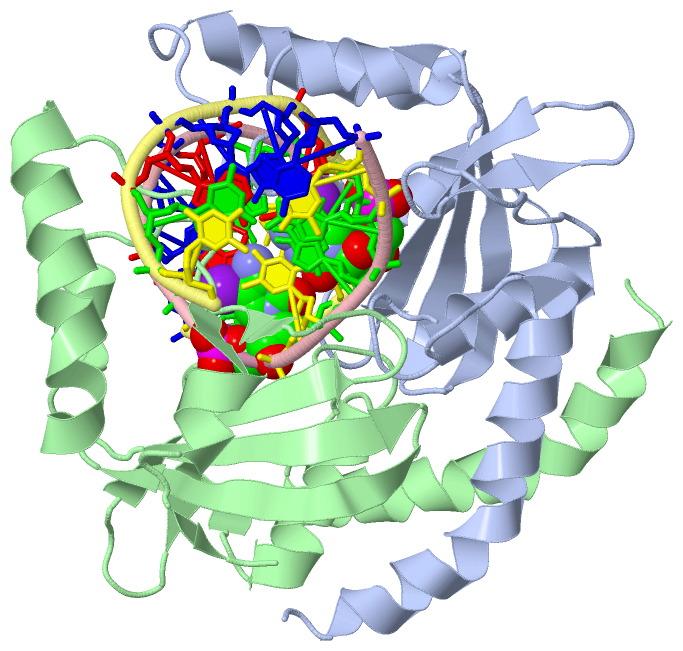
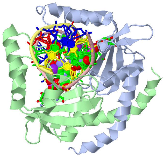
Chain A from PDB  Type:PROTEIN  Length:156
 aligned with T2P2_PROHU | P23657 from UniProtKB/Swiss-Prot  Length:157

    Alignment length:156
                                    11        21        31        41        51        61        71        81        91       101       111       121       131       141       151      
           T2P2_PROHU     2 SHPDLNKLLELWPHIQEYQDLALKHGINDIFQDNGGKLLQVLLITGLTVLPGREGNDAVDNAGQEYELKSINIDLTKGFSTHHHMNPVIIAKYRQVPWIFAIYRGIAIEAIYRLEPKDLEFYYDKWERKWYSDGHKDINNPKIPVKYVMEHGTKIY 157
               SCOP domains d2pvia_ A: Restriction endonuclease PvuII                                                                                                                    SCOP domains
               CATH domains 2pviA00 A:2-157 PVUII Endonuclease, subunit A                                                                                                                CATH domains
               Pfam domains ------------------------------------------------------------------------------------------------------------------------------------------------------------ Pfam domains
         Sec.struct. author ..hhhhhhhhhhhhhhhhhhhhhhh.........hhhhhhhhhhh...........eee.....eeeeeeee.....ee.......hhhhhhhh...eeeeeee..eeeeeeee.hhhhhhhhhhhhhhhh...........eehhhhhhh.ee.. Sec.struct. author
                 SAPs(SNPs) ------------------------------------------------------------------------------------------------------------------------------------------------------------ SAPs(SNPs)
                    PROSITE ------------------------------------------------------------------------------------------------------------------------------------------------------------ PROSITE
                 Transcript ------------------------------------------------------------------------------------------------------------------------------------------------------------ Transcript
                 2pvi A   2 SHPDLNKLLELWPHIQEYQDLALKHGINDIFQDNGGKLLQVLLITGLTVLPGRAGNDAVDNAGQEYELKSINIDLTKGFSTHHHMNPVIIAKARQVPWIFAIYRGIAIEAIYRLEPKDLEFYYDKWERKWYSDGHKDINNPKIPVKYVMEHGTKIY 157
                                    11        21        31        41        51        61        71        81        91       101       111       121       131       141       151      

Chain B from PDB  Type:PROTEIN  Length:156
 aligned with T2P2_PROHU | P23657 from UniProtKB/Swiss-Prot  Length:157

    Alignment length:156
                                    11        21        31        41        51        61        71        81        91       101       111       121       131       141       151      
           T2P2_PROHU     2 SHPDLNKLLELWPHIQEYQDLALKHGINDIFQDNGGKLLQVLLITGLTVLPGREGNDAVDNAGQEYELKSINIDLTKGFSTHHHMNPVIIAKYRQVPWIFAIYRGIAIEAIYRLEPKDLEFYYDKWERKWYSDGHKDINNPKIPVKYVMEHGTKIY 157
               SCOP domains d2pvib_ B: Restriction endonuclease PvuII                                                                                                                    SCOP domains
               CATH domains 2pviB00 B:2-157 PVUII Endonuclease, subunit A                                                                                                                CATH domains
           Pfam domains (1) -Endonuc-PvuII-2pviB01 B:3-157                                                                                                                               Pfam domains (1)
           Pfam domains (2) -Endonuc-PvuII-2pviB02 B:3-157                                                                                                                               Pfam domains (2)
         Sec.struct. author ..hhhhhhhhhhhhhhhhhhhhhhh.........hhhhhhhhhhh..ee.......eee.....eeeeeeee.....ee.......hhhhhh.....eeeeeee..eeeeeeee.hhhhhhhhhhhhhhhhhh.........eehhhhh...ee.. Sec.struct. author
                 SAPs(SNPs) ------------------------------------------------------------------------------------------------------------------------------------------------------------ SAPs(SNPs)
                    PROSITE ------------------------------------------------------------------------------------------------------------------------------------------------------------ PROSITE
                 Transcript ------------------------------------------------------------------------------------------------------------------------------------------------------------ Transcript
                 2pvi B   2 SHPDLNKLLELWPHIQEYQDLALKHGINDIFQDNGGKLLQVLLITGLTVLPGRAGNDAVDNAGQEYELKSINIDLTKGFSTHHHMNPVIIAKARQVPWIFAIYRGIAIEAIYRLEPKDLEFYYDKWERKWYSDGHKDINNPKIPVKYVMEHGTKIY 157
                                    11        21        31        41        51        61        71        81        91       101       111       121       131       141       151      

Chain C from PDB  Type:DNA  Length:13
                                             
                 2pvi C   2 TGACCAGcTGGTC  14
                                   |11   
                                   9-C38 

Chain D from PDB  Type:DNA  Length:13
                                             
                 2pvi D   2 TGACCAGcTGGTC  14
                                   |11   
                                   9-C38 

   Legend:   → Mismatch (orange background)
  - → Gap (green background, '-', border residues have a numbering label)
    → Modified Residue (blue background, lower-case, 'x' indicates undefined single-letter code, labelled with number + name)
  x → Chemical Group (purple background, 'x', labelled with number + name, e.g. ACE or NH2)
  extra numbering lines below/above indicate numbering irregularities and modified residue names etc., number ends below/above '|'

 Classification and Annotation

(-) SCOP Domains  (1, 2)

Asymmetric/Biological Unit

(-) CATH Domains  (1, 2)

Asymmetric/Biological Unit
(-)
Class: Alpha Beta (26913)

(-) Pfam Domains  (1, 2)

Asymmetric/Biological Unit

(-) Gene Ontology  (7, 7)

Asymmetric/Biological Unit(hide GO term definitions)
Chain A,B   (T2P2_PROHU | P23657)
molecular function
    GO:0009036    Type II site-specific deoxyribonuclease activity    Catalysis of the endonucleolytic cleavage of DNA to give specific double-stranded fragments with terminal 5'-phosphates and 3' hydroxyls. Cleavage is dependent on the presence in the DNA of a specific recognition site; cleavage occurs at or very near this recognition site.
    GO:0004519    endonuclease activity    Catalysis of the hydrolysis of ester linkages within nucleic acids by creating internal breaks.
    GO:0016787    hydrolase activity    Catalysis of the hydrolysis of various bonds, e.g. C-O, C-N, C-C, phosphoric anhydride bonds, etc. Hydrolase is the systematic name for any enzyme of EC class 3.
    GO:0046872    metal ion binding    Interacting selectively and non-covalently with any metal ion.
    GO:0004518    nuclease activity    Catalysis of the hydrolysis of ester linkages within nucleic acids.
biological process
    GO:0009307    DNA restriction-modification system    A defense process found in many bacteria and archaea that protects the organism from invading foreign DNA by cleaving it with a restriction endonuclease. The organism's own DNA is protected by methylation of a specific nucleotide, which occurs immediately following replication, in the same target site as the restriction enzyme.
    GO:0090305    nucleic acid phosphodiester bond hydrolysis    The nucleic acid metabolic process in which the phosphodiester bonds between nucleotides are cleaved by hydrolysis.

 Visualization

(-) Interactive Views

Asymmetric/Biological Unit
  Complete Structure
    Jena3D(integrated viewing of ligand, site, SAP, PROSITE, SCOP information)
    WebMol | AstexViewer[tm]@PDBe
(Java Applets, require no local installation except for Java; loading may be slow)
    STRAP
(Java WebStart application, automatic local installation, requires Java; full application with system access!)
    RasMol
(require local installation)
    Molscript (VRML)
(requires installation of a VRML viewer; select preferred view via VRML and generate a mono or stereo PDF format file)
 
  Ligands, Modified Residues, Ions
    C38  [ RasMol | Jena3D ]  +environment [ RasMol | Jena3D ]
 
  Sites
    CAA  [ RasMol ]  +environment [ RasMol ]
    CAB  [ RasMol ]  +environment [ RasMol ]
 
  Cis Peptide Bonds
(no "Cis Peptide Bonds" information available for 2pvi)
 

(-) Still Images

Jmol
  protein: cartoon or spacefill or dots and stick; nucleic acid: cartoon and stick; ligands: spacefill; active site: stick
Molscript
  protein, nucleic acid: cartoon; ligands: spacefill; active site: ball and stick

 Databases and Analysis Tools

(-) Databases

Access by PDB/NDB ID
  2pvi
    Family and Domain InformationProDom | SYSTERS
    General Structural InformationGlycoscienceDB | MMDB | NDB | OCA | PDB | PDBe | PDBj | PDBsum | PDBWiki | PQS | PROTEOPEDIA
    Orientation in MembranesOPM
    Protein SurfaceSURFACE
    Secondary StructureDSSP (structure derived) | HSSP (homology derived)
    Structural GenomicsGeneCensus
    Structural NeighboursCE | VAST
    Structure ClassificationCATH | Dali | SCOP
    Validation and Original DataBMRB Data View | BMRB Restraints Grid | EDS | PROCHECK | RECOORD | WHAT_CHECK
 
Access by UniProt ID/Accession number
  T2P2_PROHU | P23657
    Comparative Protein Structure ModelsModBase
    Genomic InformationEnsembl
    Protein-protein InteractionDIP
    Sequence, Family and Domain InformationInterPro | Pfam | SMART | UniProtKB/SwissProt
 
Access by Enzyme Classificator   (EC Number)
  3.1.21.4
    General Enzyme InformationBRENDA | EC-PDB | Enzyme | IntEnz
    PathwayKEGG | MetaCyc
 
Access by Disease Identifier   (MIM ID)
  (no 'MIM ID' available)
    Disease InformationOMIM
 
Access by GenAge ID
  (no 'GenAge ID' available)
    Age Related InformationGenAge

(-) Analysis Tools

Access by PDB/NDB ID
    Domain InformationXDom
    Interatomic Contacts of Structural UnitsCSU
    Ligand-protein ContactsLPC
    Protein CavitiescastP
    Sequence and Secondary StructurePDBCartoon
    Structure AlignmentSTRAP(Java WebStart application, automatic local installation, requires Java; full application with system access!)
    Structure and Sequence BrowserSTING
 
Access by UniProt ID/Accession number
  T2P2_PROHU | P23657
    Protein Disorder PredictionDisEMBL | FoldIndex | GLOBPLOT (for more information see DisProt)

 Related Entries

(-) Entries Sharing at Least One Protein Chain (UniProt ID)

UniProtKB/Swiss-Prot
        T2P2_PROHU | P236571eyu 1f0o 1h56 1k0z 1ni0 1pvi 1pvu 3ksk 3pvi

(-) Related Entries Specified in the PDB File

(no "Related Entries Specified in the PDB File" available for 2PVI)