Show PDB file:   
         Plain Text   HTML   (compressed file size)
QuickSearch:   
by PDB,NDB,UniProt,PROSITE Code or Search Term(s)  
(-)Asym.Unit - manually
(-)Asymmetric Unit
(-)Biological Unit 1
(-)Biological Unit 2
collapse expand < >
Image Asym.Unit - manually
Asym.Unit - manually  (Jmol Viewer)
Image Asymmetric Unit
Asymmetric Unit  (Jmol Viewer)
Image Biological Unit 1
Biological Unit 1  (Jmol Viewer)
Image Biological Unit 2
Biological Unit 2  (Jmol Viewer)

(-) Description

Title :  STRUCTURE OF THE Y94F MUTANT OF THE RESTRICTION ENDONUCLEASE PVUII
 
Authors :  P. A. Tucker, M. Kokkinidis, S. Nicolaki, D. Kotsifaki
Date :  20 Dec 02  (Deposition) - 04 Feb 03  (Release) - 24 Feb 09  (Revision)
Method :  X-RAY DIFFRACTION
Resolution :  2.50
Chains :  Asym. Unit :  A,B,C
Biol. Unit 1:  A  (2x)
Biol. Unit 2:  B,C  (1x)
Keywords :  Restriction Endonuclease, Pvuii, Point Mutation, Hydrolase (Keyword Search: [Gene Ontology, PubMed, Web (Google))
 
Reference :  P. A. Tucker, M. Kokkinidis, S. Nicolaki, D. Kotsifaki
Structure Of The Y94F Mutant Of The Restriction Endonuclease Pvuii
To Be Published
PubMed: search
(for further references see the PDB file header)

(-) Compounds

Molecule 1 - TYPE II RESTRICTION ENZYME PVUII
    ChainsA, B, C
    EC Number3.1.21.4
    EngineeredYES
    Expression SystemESCHERICHIA COLI
    Expression System PlasmidN-A
    Expression System StrainN/A
    Expression System Taxid562
    Expression System Vector TypeN/A
    GenePVUIIR
    MutationYES
    Organism ScientificPROTEUS VULGARIS
    Organism Taxid585
    SynonymENDONUCLEASE PVUII, R.PVUII

 Structural Features

(-) Chains, Units

  123
Asymmetric Unit ABC
Biological Unit 1 (2x)A  
Biological Unit 2 (1x) BC

Summary Information (see also Sequences/Alignments below)

(-) Ligands, Modified Residues, Ions  (0, 0)

(no "Ligand,Modified Residues,Ions" information available for 1NI0)

(-) Sites  (0, 0)

(no "Site" information available for 1NI0)

(-) SS Bonds  (0, 0)

(no "SS Bond" information available for 1NI0)

(-) Cis Peptide Bonds  (0, 0)

(no "Cis Peptide Bond" information available for 1NI0)

 Sequence-Structure Mapping

(-) SAPs(SNPs)/Variants  (0, 0)

(no "SAP(SNP)/Variant" information available for 1NI0)

(-) PROSITE Motifs  (0, 0)

(no "PROSITE Motif" information available for 1NI0)

(-) Exons   (0, 0)

(no "Exon" information available for 1NI0)

(-) Sequences/Alignments

Asymmetric Unit
   Reformat: Number of residues per line =  ('0' or empty: single-line sequence representation)
  Number of residues per labelling interval =   
  UniProt sequence: complete  aligned part    
   Show mapping: SCOP domains CATH domains Pfam domains Secondary structure (by author)
SAPs(SNPs) PROSITE motifs Exons
(details for a mapped element are shown in a popup box when the mouse pointer rests over it)
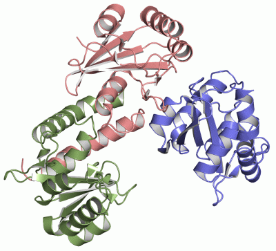
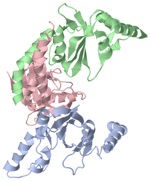
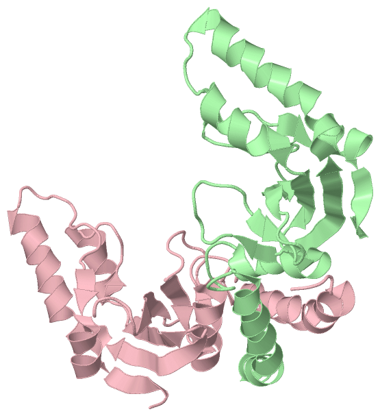
Chain A from PDB  Type:PROTEIN  Length:157
 aligned with T2P2_PROHU | P23657 from UniProtKB/Swiss-Prot  Length:157

    Alignment length:157
                                                                                                                                                                                    157  
                                    12        22        32        42        52        62        72        82        92       102       112       122       132       142       152    |  
           T2P2_PROHU     3 HPDLNKLLELWPHIQEYQDLALKHGINDIFQDNGGKLLQVLLITGLTVLPGREGNDAVDNAGQEYELKSINIDLTKGFSTHHHMNPVIIAKYRQVPWIFAIYRGIAIEAIYRLEPKDLEFYYDKWERKWYSDGHKDINNPKIPVKYVMEHGTKIY--   -
               SCOP domains d1ni0a_ A: Restriction endonuclease PvuII                                                                                                                     SCOP domains
               CATH domains 1ni0A00 A:3-159 PVUII Endonuclease, subunit A                                                                                                                 CATH domains
               Pfam domains ------------------------------------------------------------------------------------------------------------------------------------------------------------- Pfam domains
         Sec.struct. author hhhhhhhhhhhhhhhhhhhhhhhh........hhhhhhhhhhhhh.ee........ee.....eeeeeeee.....ee......hhhhhhhhh...eeeeeee..eeeeeeee.hhhhhhhhhhhhhhhhh..........eehhhhhhhheeeee. Sec.struct. author
                 SAPs(SNPs) ------------------------------------------------------------------------------------------------------------------------------------------------------------- SAPs(SNPs)
                    PROSITE ------------------------------------------------------------------------------------------------------------------------------------------------------------- PROSITE
                 Transcript ------------------------------------------------------------------------------------------------------------------------------------------------------------- Transcript
                 1ni0 A   3 HPDLNKLLELWPHIQEYQDLALKHGINDIFQDNGGKLLQVLLITGLTVLPGREGNDAVDNAGQEYELKSINIDLTKGFSTHHHMNPVIIAKFRQVPWIFAIYRGIAIEAIYRLEPKDLEFYYDKWERKWYSDGHKDINNPKIPVKYVMEHGTKIYAA 159
                                    12        22        32        42        52        62        72        82        92       102       112       122       132       142       152       

Chain B from PDB  Type:PROTEIN  Length:157
 aligned with T2P2_PROHU | P23657 from UniProtKB/Swiss-Prot  Length:157

    Alignment length:157
                                                                                                                                                                                    157  
                                    12        22        32        42        52        62        72        82        92       102       112       122       132       142       152    |  
           T2P2_PROHU     3 HPDLNKLLELWPHIQEYQDLALKHGINDIFQDNGGKLLQVLLITGLTVLPGREGNDAVDNAGQEYELKSINIDLTKGFSTHHHMNPVIIAKYRQVPWIFAIYRGIAIEAIYRLEPKDLEFYYDKWERKWYSDGHKDINNPKIPVKYVMEHGTKIY--   -
               SCOP domains d1ni0b_ B: Restriction endonuclease PvuII                                                                                                                     SCOP domains
               CATH domains 1ni0B00 B:3-159 PVUII Endonuclease, subunit A                                                                                                                 CATH domains
               Pfam domains ------------------------------------------------------------------------------------------------------------------------------------------------------------- Pfam domains
         Sec.struct. author hhhhhhhhhhhhhhhhhhhhhhhh........hhhhhhhhhhhhh.ee........ee.....eeeeeeee.....ee....eehhhhhhhhhh..eeeeeee..eeeeeeeehhhhhhhhhhhhhhhhhhh....ee...eehhhhhhhheeeee. Sec.struct. author
                 SAPs(SNPs) ------------------------------------------------------------------------------------------------------------------------------------------------------------- SAPs(SNPs)
                    PROSITE ------------------------------------------------------------------------------------------------------------------------------------------------------------- PROSITE
                 Transcript ------------------------------------------------------------------------------------------------------------------------------------------------------------- Transcript
                 1ni0 B   3 HPDLNKLLELWPHIQEYQDLALKHGINDIFQDNGGKLLQVLLITGLTVLPGREGNDAVDNAGQEYELKSINIDLTKGFSTHHHMNPVIIAKFRQVPWIFAIYRGIAIEAIYRLEPKDLEFYYDKWERKWYSDGHKDINNPKIPVKYVMEHGTKIYAA 159
                                    12        22        32        42        52        62        72        82        92       102       112       122       132       142       152       

Chain C from PDB  Type:PROTEIN  Length:158
 aligned with T2P2_PROHU | P23657 from UniProtKB/Swiss-Prot  Length:157

    Alignment length:158
                                                                                                                                                                                     157  
                                    11        21        31        41        51        61        71        81        91       101       111       121       131       141       151     |  
           T2P2_PROHU     2 SHPDLNKLLELWPHIQEYQDLALKHGINDIFQDNGGKLLQVLLITGLTVLPGREGNDAVDNAGQEYELKSINIDLTKGFSTHHHMNPVIIAKYRQVPWIFAIYRGIAIEAIYRLEPKDLEFYYDKWERKWYSDGHKDINNPKIPVKYVMEHGTKIY--   -
               SCOP domains d1ni0c_ C: Restriction endonuclease PvuII                                                                                                                      SCOP domains
               CATH domains 1ni0C00 C:2-159 PVUII Endonuclease, subunit A                                                                                                                  CATH domains
           Pfam domains (1) -Endonuc-PvuII-1ni0C01 C:3-157                                                                                                                              -- Pfam domains (1)
           Pfam domains (2) -Endonuc-PvuII-1ni0C02 C:3-157                                                                                                                              -- Pfam domains (2)
           Pfam domains (3) -Endonuc-PvuII-1ni0C03 C:3-157                                                                                                                              -- Pfam domains (3)
         Sec.struct. author ...hhhhhhhhhhhhhhhhhhhhhh........hhhhhhhhhhhhh.ee.......eee.....eeeeeeee......ee.....hhhhhhhhh...eeeeeee..eeeeeeeehhhhhhhhhhhhhhhhhh.........eehhhhhhhhheeee.. Sec.struct. author
                 SAPs(SNPs) -------------------------------------------------------------------------------------------------------------------------------------------------------------- SAPs(SNPs)
                    PROSITE -------------------------------------------------------------------------------------------------------------------------------------------------------------- PROSITE
                 Transcript -------------------------------------------------------------------------------------------------------------------------------------------------------------- Transcript
                 1ni0 C   2 SHPDLNKLLELWPHIQEYQDLALKHGINDIFQDNGGKLLQVLLITGLTVLPGREGNDAVDNAGQEYELKSINIDLTKGFSTHHHMNPVIIAKFRQVPWIFAIYRGIAIEAIYRLEPKDLEFYYDKWERKWYSDGHKDINNPKIPVKYVMEHGTKIYAA 159
                                    11        21        31        41        51        61        71        81        91       101       111       121       131       141       151        

   Legend:   → Mismatch (orange background)
  - → Gap (green background, '-', border residues have a numbering label)
    → Modified Residue (blue background, lower-case, 'x' indicates undefined single-letter code, labelled with number + name)
  x → Chemical Group (purple background, 'x', labelled with number + name, e.g. ACE or NH2)
  extra numbering lines below/above indicate numbering irregularities and modified residue names etc., number ends below/above '|'

 Classification and Annotation

(-) SCOP Domains  (1, 3)

Asymmetric Unit

(-) CATH Domains  (1, 3)

Asymmetric Unit
(-)
Class: Alpha Beta (26913)

(-) Pfam Domains  (1, 3)

Asymmetric Unit

(-) Gene Ontology  (7, 7)

Asymmetric Unit(hide GO term definitions)
Chain A,B,C   (T2P2_PROHU | P23657)
molecular function
    GO:0009036    Type II site-specific deoxyribonuclease activity    Catalysis of the endonucleolytic cleavage of DNA to give specific double-stranded fragments with terminal 5'-phosphates and 3' hydroxyls. Cleavage is dependent on the presence in the DNA of a specific recognition site; cleavage occurs at or very near this recognition site.
    GO:0004519    endonuclease activity    Catalysis of the hydrolysis of ester linkages within nucleic acids by creating internal breaks.
    GO:0016787    hydrolase activity    Catalysis of the hydrolysis of various bonds, e.g. C-O, C-N, C-C, phosphoric anhydride bonds, etc. Hydrolase is the systematic name for any enzyme of EC class 3.
    GO:0046872    metal ion binding    Interacting selectively and non-covalently with any metal ion.
    GO:0004518    nuclease activity    Catalysis of the hydrolysis of ester linkages within nucleic acids.
biological process
    GO:0009307    DNA restriction-modification system    A defense process found in many bacteria and archaea that protects the organism from invading foreign DNA by cleaving it with a restriction endonuclease. The organism's own DNA is protected by methylation of a specific nucleotide, which occurs immediately following replication, in the same target site as the restriction enzyme.
    GO:0090305    nucleic acid phosphodiester bond hydrolysis    The nucleic acid metabolic process in which the phosphodiester bonds between nucleotides are cleaved by hydrolysis.

 Visualization

(-) Interactive Views

Asymmetric Unit
  Complete Structure
    Jena3D(integrated viewing of ligand, site, SAP, PROSITE, SCOP information)
    WebMol | AstexViewer[tm]@PDBe
(Java Applets, require no local installation except for Java; loading may be slow)
    STRAP
(Java WebStart application, automatic local installation, requires Java; full application with system access!)
    RasMol
(require local installation)
    Molscript (VRML)
(requires installation of a VRML viewer; select preferred view via VRML and generate a mono or stereo PDF format file)
 
  Ligands, Modified Residues, Ions
(no "Ligands, Modified Residues, Ions" information available for 1ni0)
 
  Sites
(no "Sites" information available for 1ni0)
 
  Cis Peptide Bonds
(no "Cis Peptide Bonds" information available for 1ni0)
 
Biological Units
  Complete Structure
    Biological Unit 1  [ Jena3D ]
    Biological Unit 2  [ Jena3D ]

(-) Still Images

Jmol
  protein: cartoon or spacefill or dots and stick; nucleic acid: cartoon and stick; ligands: spacefill; active site: stick
Molscript
  protein, nucleic acid: cartoon; ligands: spacefill; active site: ball and stick

 Databases and Analysis Tools

(-) Databases

Access by PDB/NDB ID
  1ni0
    Family and Domain InformationProDom | SYSTERS
    General Structural InformationGlycoscienceDB | MMDB | NDB | OCA | PDB | PDBe | PDBj | PDBsum | PDBWiki | PQS | PROTEOPEDIA
    Orientation in MembranesOPM
    Protein SurfaceSURFACE
    Secondary StructureDSSP (structure derived) | HSSP (homology derived)
    Structural GenomicsGeneCensus
    Structural NeighboursCE | VAST
    Structure ClassificationCATH | Dali | SCOP
    Validation and Original DataBMRB Data View | BMRB Restraints Grid | EDS | PROCHECK | RECOORD | WHAT_CHECK
 
Access by UniProt ID/Accession number
  T2P2_PROHU | P23657
    Comparative Protein Structure ModelsModBase
    Genomic InformationEnsembl
    Protein-protein InteractionDIP
    Sequence, Family and Domain InformationInterPro | Pfam | SMART | UniProtKB/SwissProt
 
Access by Enzyme Classificator   (EC Number)
  3.1.21.4
    General Enzyme InformationBRENDA | EC-PDB | Enzyme | IntEnz
    PathwayKEGG | MetaCyc
 
Access by Disease Identifier   (MIM ID)
  (no 'MIM ID' available)
    Disease InformationOMIM
 
Access by GenAge ID
  (no 'GenAge ID' available)
    Age Related InformationGenAge

(-) Analysis Tools

Access by PDB/NDB ID
    Domain InformationXDom
    Interatomic Contacts of Structural UnitsCSU
    Ligand-protein ContactsLPC
    Protein CavitiescastP
    Sequence and Secondary StructurePDBCartoon
    Structure AlignmentSTRAP(Java WebStart application, automatic local installation, requires Java; full application with system access!)
    Structure and Sequence BrowserSTING
 
Access by UniProt ID/Accession number
  T2P2_PROHU | P23657
    Protein Disorder PredictionDisEMBL | FoldIndex | GLOBPLOT (for more information see DisProt)

 Related Entries

(-) Entries Sharing at Least One Protein Chain (UniProt ID)

UniProtKB/Swiss-Prot
        T2P2_PROHU | P236571eyu 1f0o 1h56 1k0z 1pvi 1pvu 2pvi 3ksk 3pvi

(-) Related Entries Specified in the PDB File

1pvu NATIVE STRUCTURE OF PVUII
3pvi STRUCTURE OF PVUII IN COMPLEX WITH DNA