| molecular function |
|---|
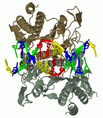
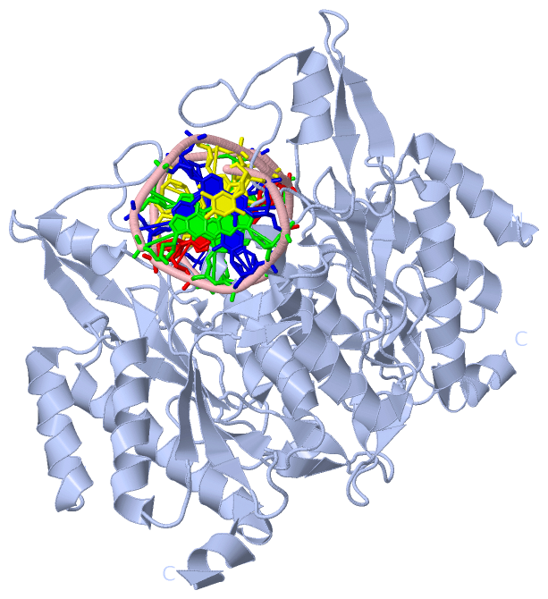
| | GO:0003677 | | DNA binding | | Any molecular function by which a gene product interacts selectively and non-covalently with DNA (deoxyribonucleic acid). |
| | GO:0009036 | | Type II site-specific deoxyribonuclease activity | | Catalysis of the endonucleolytic cleavage of DNA to give specific double-stranded fragments with terminal 5'-phosphates and 3' hydroxyls. Cleavage is dependent on the presence in the DNA of a specific recognition site; cleavage occurs at or very near this recognition site. |
| | GO:0004519 | | endonuclease activity | | Catalysis of the hydrolysis of ester linkages within nucleic acids by creating internal breaks. |
| | GO:0016787 | | hydrolase activity | | Catalysis of the hydrolysis of various bonds, e.g. C-O, C-N, C-C, phosphoric anhydride bonds, etc. Hydrolase is the systematic name for any enzyme of EC class 3. |
| | GO:0000287 | | magnesium ion binding | | Interacting selectively and non-covalently with magnesium (Mg) ions. |
| | GO:0046872 | | metal ion binding | | Interacting selectively and non-covalently with any metal ion. |
| | GO:0004518 | | nuclease activity | | Catalysis of the hydrolysis of ester linkages within nucleic acids. |
| biological process |
|---|
| | GO:0009307 | | DNA restriction-modification system | | A defense process found in many bacteria and archaea that protects the organism from invading foreign DNA by cleaving it with a restriction endonuclease. The organism's own DNA is protected by methylation of a specific nucleotide, which occurs immediately following replication, in the same target site as the restriction enzyme. |
| | GO:0090305 | | nucleic acid phosphodiester bond hydrolysis | | The nucleic acid metabolic process in which the phosphodiester bonds between nucleotides are cleaved by hydrolysis. |
 Description