|
|
|
|
 Description
|
|
Compounds
|
||||||||||||||||||||||||||||||||||||||||||||||||||||||||||||||||||||
Chains, Units
Summary Information (see also Sequences/Alignments below) |
Ligands, Modified Residues, Ions (1, 1)
Asymmetric Unit (1, 1)
|
Sites (1, 1)
Asymmetric Unit (1, 1)
|
SS Bonds (0, 0)| (no "SS Bond" information available for 1QPS) |
Cis Peptide Bonds (0, 0)| (no "Cis Peptide Bond" information available for 1QPS) |
SAPs(SNPs)/Variants (0, 0)| (no "SAP(SNP)/Variant" information available for 1QPS) |
PROSITE Motifs (0, 0)| (no "PROSITE Motif" information available for 1QPS) |
Exons (0, 0)| (no "Exon" information available for 1QPS) |
Sequences/Alignments
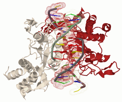
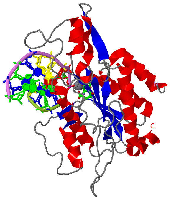
Asymmetric UnitChain A from PDB Type:PROTEIN Length:261 aligned with T2E1_ECOLX | P00642 from UniProtKB/Swiss-Prot Length:277 Alignment length:261 26 36 46 56 66 76 86 96 106 116 126 136 146 156 166 176 186 196 206 216 226 236 246 256 266 276 T2E1_ECOLX 17 SQGVIGIFGDYAKAHDLAVGEVSKLVKKALSNEYPQLSFRYRDSIKKTEINEALKKIDPDLGGTLFVSNSSIKPDGGIVEVKDDYGEWRVVLVAEAKHQGKDIINIRNGLLVGKRGDQDLMAAGNAIERSHKNISEIANFMLSESHFPYVLFLEGSNFLTENISITRPDGRVVNLEYNSGILNRLDRLTAANYGMPINSNLCINKFVNHKDKSIMLQAASIYTQGDGREWDSKIMFEIMFDISTTSLRVLGRDLFEQLTSK 277 SCOP domains d1qpsa_ A: Restriction endonuclease EcoRI SCOP domains CATH domains 1qpsA00 A:17-277 Eco RI Endonuclease, subunit A CATH domains Pfam domains EcoRI-1qpsA01 A:17-273 ---- Pfam domains SAPs(SNPs) --------------------------------------------------------------------------------------------------------------------------------------------------------------------------------------------------------------------------------------------------------------------- SAPs(SNPs) PROSITE --------------------------------------------------------------------------------------------------------------------------------------------------------------------------------------------------------------------------------------------------------------------- PROSITE Transcript --------------------------------------------------------------------------------------------------------------------------------------------------------------------------------------------------------------------------------------------------------------------- Transcript 1qps A 17 SQGVIGIFGDYAKAHDLAVGEVSKLVKKALSNEYPQLSFRYRDSIKKTEINEALKKIDPDLGGTLFVSNSSIKPDGGIVEVKDDYGEWRVVLVAEAKHQGKDIINIRNGLLVGKRGDQDLMAAGNAIERSHKNISEIANFMLSESHFPYVLFLEGSNFLTENISITRPDGRVVNLEYNSGILNRLDRLTAANYGMPINSNLCINKFVNHKDKSIMLQAASIYTQGDGREWDSKIMFEIMFDISTTSLRVLGRDLFEQLTSK 277 26 36 46 56 66 76 86 96 106 116 126 136 146 156 166 176 186 196 206 216 226 236 246 256 266 276
Chain M from PDB  Type:DNA  Length:5

                 1qps M   1 TCGCG   5
Chain N from PDB  Type:DNA  Length:8

                 1qps N   6 AATTCGCG  13
|
||||||||||||||||||||
SCOP Domains (1, 1)
Asymmetric Unit
 |
CATH Domains (1, 1)
Asymmetric Unit
|
Pfam Domains (1, 1)
Asymmetric Unit
|
Gene Ontology (9, 9)|
Asymmetric Unit(hide GO term definitions) Chain A (T2E1_ECOLX | P00642)
|
||||||||||||||||||||||||||||||||||||||||||||||||||||||||||||||||||
Interactive Views
|
||||||||||||||||||||||||||||||||||||||||||||||||||||||||||||||||||||||||||||||||||||||||||||||||||||||||||||||||||||||||||||||||||||||||
Still Images
|
||||||||||||||||
Databases
|
||||||||||||||||||||||||||||||||||||||||||||||||||||||||||||||||||||||||||||||||||||||||||||||||||||||||||||||||||||||||||||||||||||||||||||||||||||||||||||||||
Analysis Tools
|
|||||||||||||||||||||||||||||||||||||||||||||||||||||||||||||
Entries Sharing at Least One Protein Chain (UniProt ID)
Related Entries Specified in the PDB File
|
|