Show PDB file:   
         Plain Text   HTML   (compressed file size)
QuickSearch:   
by PDB,NDB,UniProt,PROSITE Code or Search Term(s)  
(-)NMR Structure - manually
(-)NMR Structure - model 1
(-)NMR Structure - all models
collapse expand < >
Image NMR Structure - manually
NMR Structure - manually  (Jmol Viewer)
Image NMR Structure - model 1
NMR Structure - model 1  (Jmol Viewer)
Image NMR Structure - all models
NMR Structure - all models  (Jmol Viewer)

(-) Description

Title :  SOLUTION STRUCTURE OF THE RGS DOMAIN FROM HUMAN RGS18
 
Authors :  V. A. Higman, M. Leidert, J. Bray, J. Elkins, M. Soundararajan, D. A. Doy C. Gileadi, C. Phillips, G. Schoch, X. Yang, C. Brockmann, P. Schmiede A. Diehl, M. Sundstrom, C. Arrowsmith, J. Weigelt, A. Edwards, H. Osch L. J. Ball, Structural Genomics Consortium (Sgc)
Date :  16 Feb 07  (Deposition) - 27 Feb 07  (Release) - 15 Aug 12  (Revision)
Method :  SOLUTION NMR
Resolution :  NOT APPLICABLE
Chains :  NMR Structure  :  A  (20x)
NMR Structure *:  A  (1x)
Keywords :  Signaling Protein, Structural Genomics, Structural Genomics Consortium, Sgc (Keyword Search: [Gene Ontology, PubMed, Web (Google))
 
Reference :  M. Soundararajan, F. S. Willard, A. J. Kimple, A. P. Turnbull, L. J. Ball, G. A. Schoch, C. Gileadi, O. Y. Fedorov, E. F. Dowler, V. A. Higman, S. Q. Hutsell, M. Sundstrom, D. A. Doyle, D. P. Siderovski
Structural Diversity In The Rgs Domain And Its Interaction With Heterotrimeric G Protein Alpha-Subunits.
Proc. Natl. Acad. Sci. Usa V. 105 6457 2008
PubMed-ID: 18434541  |  Reference-DOI: 10.1073/PNAS.0801508105
(for further references see the PDB file header)

(-) Compounds

Molecule 1 - REGULATOR OF G-PROTEIN SIGNALING 18
    ChainsA
    EngineeredYES
    Expression SystemESCHERICHIA COLI
    Expression System StrainBL21 (DE3) - ROSETTA
    Expression System Taxid562
    FragmentRGS DOMAIN, RESIDUES 75-223
    GeneRGS18, RGS13
    Organism CommonHUMAN
    Organism ScientificHOMO SAPIENS
    Organism Taxid9606
    Other DetailsBL21 (DE3) STRAIN ENRICHED WITH GENES THAT ENCODE RARE TRNAS
    SynonymRGS18

 Structural Features

(-) Chains, Units

  1
NMR Structure (20x)A
NMR Structure * (1x)A

Summary Information (see also Sequences/Alignments below)

(-) Ligands, Modified Residues, Ions  (0, 0)

(no "Ligand,Modified Residues,Ions" information available for 2OWI)

(-) Sites  (0, 0)

(no "Site" information available for 2OWI)

(-) SS Bonds  (0, 0)

(no "SS Bond" information available for 2OWI)

(-) Cis Peptide Bonds  (0, 0)

(no "Cis Peptide Bond" information available for 2OWI)

 Sequence-Structure Mapping

(-) SAPs(SNPs)/Variants  (0, 0)

(no "SAP(SNP)/Variant" information available for 2OWI)

(-) PROSITE Motifs  (1, 1)

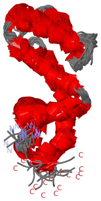
NMR Structure (1, 1)
 PROSITEUniProtKBPDB
No.IDACDescriptionIDLocationCountLocation
1RGSPS50132 RGS domain profile.RGS18_HUMAN86-202  1A:14-130
NMR Structure * (1, 1)
 PROSITEUniProtKBPDB
No.IDACDescriptionIDLocationCountLocation
1RGSPS50132 RGS domain profile.RGS18_HUMAN86-202  1A:14-130

(-) Exons   (4, 4)

NMR Structure (4, 4)
 ENSEMBLUniProtKBPDB
No.Transcript IDExonExon IDGenome LocationLengthIDLocationLengthCountLocationLength
1.1aENST000003674601aENSE00001444556chr1:192127587-192127886300RGS18_HUMAN1-40400--
1.2ENST000003674602ENSE00000995892chr1:192128350-192128451102RGS18_HUMAN40-74351A:1-22
1.3aENST000003674603aENSE00000995891chr1:192129508-19212956962RGS18_HUMAN74-95221A:2-2322
1.6bENST000003674606bENSE00000995890chr1:192150422-192150588167RGS18_HUMAN95-150561A:23-7856
1.7ENST000003674607ENSE00001444555chr1:192153427-1921549451519RGS18_HUMAN151-235851A:79-13456

(-) Sequences/Alignments

NMR Structure
   Reformat: Number of residues per line =  ('0' or empty: single-line sequence representation)
  Number of residues per labelling interval =   
  UniProt sequence: complete  aligned part    
   Show mapping: SCOP domains CATH domains Pfam domains Secondary structure (by author)
SAPs(SNPs) PROSITE motifs Exons
(details for a mapped element are shown in a popup box when the mouse pointer rests over it)
Chain A from PDB  Type:PROTEIN  Length:134
 aligned with RGS18_HUMAN | Q9NS28 from UniProtKB/Swiss-Prot  Length:235

    Alignment length:134
                                    82        92       102       112       122       132       142       152       162       172       182       192       202    
          RGS18_HUMAN    73 TRVSPEEAVKWGESFDKLLSHRDGLEAFTRFLKTEFSEENIEFWIACEDFKKSKGPQQIHLKAKAIYEKFIQTDAPKEVNLDFHTKEVITNSITQPTLHSFDAAQSRVYQLMEQDSYTRFLKSDIYLDLMEGRP 206
               SCOP domains d2owia_ A: automated matches                                                                                                           SCOP domains
               CATH domains -------------------------------------------------------------------------------------------------------------------------------------- CATH domains
               Pfam domains -------------RGS-2owiA01 A:14-129                                                                                                ----- Pfam domains
         Sec.struct. author ...hhhhhhhhh.hhhhhhhhhhhhhhhhhhhhh..hhhhhhhhhhhhhhhh..hhhhhhhhhhhhhhhh...........hhhhhhhhhhhh.......hhhhhhhhhhhhhhhhhhhhhhhhhhhhhhh... Sec.struct. author
                 SAPs(SNPs) -------------------------------------------------------------------------------------------------------------------------------------- SAPs(SNPs)
                    PROSITE -------------RGS  PDB: A:14-130 UniProt: 86-202                                                                                   ---- PROSITE
           Transcript 1 (1) 1.--------------------Exon 1.6b  PDB: A:23-78 UniProt: 95-150                 Exon 1.7  PDB: A:79-134 UniProt: 151-235 [INCOMPLETE]    Transcript 1 (1)
           Transcript 1 (2) -Exon 1.3a  PDB: A:2-23--------------------------------------------------------------------------------------------------------------- Transcript 1 (2)
                 2owi A   1 SMVSPEEAVKWGESFDKLLSHRDGLEAFTRFLKTEFSEENIEFWIACEDFKKSKGPQQIHLKAKAIYEKFIQTDAPKEVNLDFHTKEVITNSITQPTLHSFDAAQSRVYQLMEQDSYTRFLKSDIYLDLMEGRP 134
                                    10        20        30        40        50        60        70        80        90       100       110       120       130    

   Legend:   → Mismatch (orange background)
  - → Gap (green background, '-', border residues have a numbering label)
    → Modified Residue (blue background, lower-case, 'x' indicates undefined single-letter code, labelled with number + name)
  x → Chemical Group (purple background, 'x', labelled with number + name, e.g. ACE or NH2)
  extra numbering lines below/above indicate numbering irregularities and modified residue names etc., number ends below/above '|'

 Classification and Annotation

(-) SCOP Domains  (1, 1)

NMR Structure

(-) CATH Domains  (0, 0)

(no "CATH Domain" information available for 2OWI)

(-) Pfam Domains  (1, 1)

NMR Structure
(-)
Clan: RGS (17)

(-) Gene Ontology  (7, 7)

NMR Structure(hide GO term definitions)
Chain A   (RGS18_HUMAN | Q9NS28)
molecular function
    GO:0005096    GTPase activator activity    Binds to and increases the activity of a GTPase, an enzyme that catalyzes the hydrolysis of GTP.
biological process
    GO:0007186    G-protein coupled receptor signaling pathway    A series of molecular signals that proceeds with an activated receptor promoting the exchange of GDP for GTP on the alpha-subunit of an associated heterotrimeric G-protein complex. The GTP-bound activated alpha-G-protein then dissociates from the beta- and gamma-subunits to further transmit the signal within the cell. The pathway begins with receptor-ligand interaction, or for basal GPCR signaling the pathway begins with the receptor activating its G protein in the absence of an agonist, and ends with regulation of a downstream cellular process, e.g. transcription. The pathway can start from the plasma membrane, Golgi or nuclear membrane (PMID:24568158 and PMID:16902576).
    GO:0009968    negative regulation of signal transduction    Any process that stops, prevents, or reduces the frequency, rate or extent of signal transduction.
    GO:0043547    positive regulation of GTPase activity    Any process that activates or increases the activity of a GTPase.
    GO:0008277    regulation of G-protein coupled receptor protein signaling pathway    Any process that modulates the frequency, rate or extent of G-protein coupled receptor protein signaling pathway activity.
cellular component
    GO:0005737    cytoplasm    All of the contents of a cell excluding the plasma membrane and nucleus, but including other subcellular structures.
    GO:0005886    plasma membrane    The membrane surrounding a cell that separates the cell from its external environment. It consists of a phospholipid bilayer and associated proteins.

 Visualization

(-) Interactive Views

NMR Structure
  Complete Structure
    Jena3D(integrated viewing of ligand, site, SAP, PROSITE, SCOP information)
    WebMol | AstexViewer[tm]@PDBe
(Java Applets, require no local installation except for Java; loading may be slow)
    STRAP
(Java WebStart application, automatic local installation, requires Java; full application with system access!)
    RasMol
(require local installation)
    Molscript (VRML)
(requires installation of a VRML viewer; select preferred view via VRML and generate a mono or stereo PDF format file)
 
  Ligands, Modified Residues, Ions
(no "Ligands, Modified Residues, Ions" information available for 2owi)
 
  Sites
(no "Sites" information available for 2owi)
 
  Cis Peptide Bonds
(no "Cis Peptide Bonds" information available for 2owi)
 

(-) Still Images

Jmol
  protein: cartoon or spacefill or dots and stick; nucleic acid: cartoon and stick; ligands: spacefill; active site: stick
Molscript
  protein, nucleic acid: cartoon; ligands: spacefill; active site: ball and stick

 Databases and Analysis Tools

(-) Databases

Access by PDB/NDB ID
  2owi
    Family and Domain InformationProDom | SYSTERS
    General Structural InformationGlycoscienceDB | MMDB | NDB | OCA | PDB | PDBe | PDBj | PDBsum | PDBWiki | PQS | PROTEOPEDIA
    Orientation in MembranesOPM
    Protein SurfaceSURFACE
    Secondary StructureDSSP (structure derived) | HSSP (homology derived)
    Structural GenomicsGeneCensus
    Structural NeighboursCE | VAST
    Structure ClassificationCATH | Dali | SCOP
    Validation and Original DataBMRB Data View | BMRB Restraints Grid | EDS | PROCHECK | RECOORD | WHAT_CHECK
 
Access by UniProt ID/Accession number
  RGS18_HUMAN | Q9NS28
    Comparative Protein Structure ModelsModBase
    Genomic InformationEnsembl
    Protein-protein InteractionDIP
    Sequence, Family and Domain InformationInterPro | Pfam | SMART | UniProtKB/SwissProt
 
Access by Enzyme Classificator   (EC Number)
  (no 'Enzyme Classificator' available)
    General Enzyme InformationBRENDA | EC-PDB | Enzyme | IntEnz
    PathwayKEGG | MetaCyc
 
Access by Disease Identifier   (MIM ID)
  (no 'MIM ID' available)
    Disease InformationOMIM
 
Access by GenAge ID
  (no 'GenAge ID' available)
    Age Related InformationGenAge

(-) Analysis Tools

Access by PDB/NDB ID
    Domain InformationXDom
    Interatomic Contacts of Structural UnitsCSU
    Ligand-protein ContactsLPC
    Protein CavitiescastP
    Sequence and Secondary StructurePDBCartoon
    Structure AlignmentSTRAP(Java WebStart application, automatic local installation, requires Java; full application with system access!)
    Structure and Sequence BrowserSTING
 
Access by UniProt ID/Accession number
  RGS18_HUMAN | Q9NS28
    Protein Disorder PredictionDisEMBL | FoldIndex | GLOBPLOT (for more information see DisProt)

 Related Entries

(-) Entries Sharing at Least One Protein Chain (UniProt ID)

UniProtKB/Swiss-Prot
        RGS18_HUMAN | Q9NS282dlv 2jm5

(-) Related Entries Specified in the PDB File

7106 CHEMICAL SHIFTS
2jm5 STRUCTURE CALCULATED FROM THE SAME RESTRAINTS, BUT USING A DIFFERENT REFINEMENT PROTOCOL