Show PDB file:   
         Plain Text   HTML   (compressed file size)
QuickSearch:   
by PDB,NDB,UniProt,PROSITE Code or Search Term(s)  
(-)NMR Structure - manually
(-)NMR Structure - model 1
(-)NMR Structure - all models
collapse expand < >
Image NMR Structure - manually
NMR Structure - manually  (Jmol Viewer)
Image NMR Structure - model 1
NMR Structure - model 1  (Jmol Viewer)
Image NMR Structure - all models
NMR Structure - all models  (Jmol Viewer)

(-) Description

Title :  SOLUTION STRUCTURE OF THE RGS DOMAIN OF HUMAN REGULATOR OF G-PROTEIN SIGNALING 18
 
Authors :  H. P. Zhang, T. Suetake, F. Hayashi, S. Yokoyama, Riken Structural Genomics/Proteomics Initiative (Rsgi)
Date :  20 Apr 06  (Deposition) - 17 Apr 07  (Release) - 24 Feb 09  (Revision)
Method :  SOLUTION NMR
Resolution :  NOT APPLICABLE
Chains :  NMR Structure  :  A  (20x)
Keywords :  Rgs Domain, Regulator Of G-Protein Signaling 18, Rgs18, Structural Genomics, Nppsfa, National Project On Protein Structural And Functional Analyses, Riken Structural Genomics/Proteomics Initiative, Rsgi, Signaling Protein (Keyword Search: [Gene Ontology, PubMed, Web (Google))
 
Reference :  H. P. Zhang, T. Suetake, F. Hayashi, S. Yokoyama
Solution Structure Of The Rgs Domain Of Human Regulator Of G-Protein Signaling 18
To Be Published
PubMed: search
(for further references see the PDB file header)

(-) Compounds

Molecule 1 - REGULATOR OF G-PROTEIN SIGNALING 18
    ChainsA
    EngineeredYES
    Expression System PlasmidP050627-05
    Expression System Vector TypePLASMID
    FragmentRGS DOMAIN
    GeneRGS18, RGS13
    Organism CommonHUMAN
    Organism ScientificHOMO SAPIENS
    Organism Taxid9606
    Other DetailsCELL FREE PROTEIN SYNTHESIS
    SynonymRGS18

 Structural Features

(-) Chains, Units

  
NMR Structure (20x)

Summary Information (see also Sequences/Alignments below)

(-) Ligands, Modified Residues, Ions  (0, 0)

(no "Ligand,Modified Residues,Ions" information available for 2DLV)

(-) Sites  (0, 0)

(no "Site" information available for 2DLV)

(-) SS Bonds  (0, 0)

(no "SS Bond" information available for 2DLV)

(-) Cis Peptide Bonds  (0, 0)

(no "Cis Peptide Bond" information available for 2DLV)

 Sequence-Structure Mapping

(-) SAPs(SNPs)/Variants  (0, 0)

(no "SAP(SNP)/Variant" information available for 2DLV)

(-) PROSITE Motifs  (1, 1)

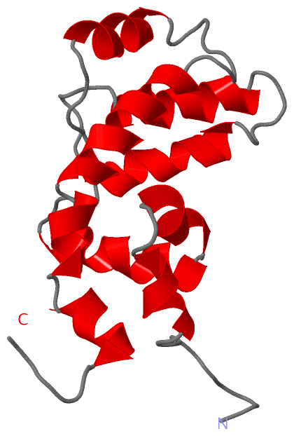
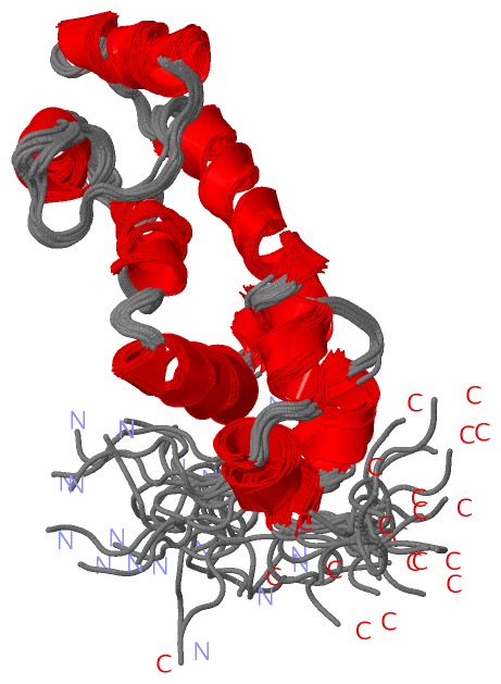
NMR Structure (1, 1)
 PROSITEUniProtKBPDB
No.IDACDescriptionIDLocationCountLocation
1RGSPS50132 RGS domain profile.RGS18_HUMAN86-202  1A:18-134

(-) Exons   (5, 5)

NMR Structure (5, 5)
 ENSEMBLUniProtKBPDB
No.Transcript IDExonExon IDGenome LocationLengthIDLocationLengthCountLocationLength
1.1aENST000003674601aENSE00001444556chr1:192127587-192127886300RGS18_HUMAN1-40401A:1-22
1.2ENST000003674602ENSE00000995892chr1:192128350-192128451102RGS18_HUMAN40-74351A:3-75
1.3aENST000003674603aENSE00000995891chr1:192129508-19212956962RGS18_HUMAN74-95221A:8-2720
1.6bENST000003674606bENSE00000995890chr1:192150422-192150588167RGS18_HUMAN95-150561A:27-8256
1.7ENST000003674607ENSE00001444555chr1:192153427-1921549451519RGS18_HUMAN151-235851A:83-140 (gaps)67

(-) Sequences/Alignments

NMR Structure
   Reformat: Number of residues per line =  ('0' or empty: single-line sequence representation)
  Number of residues per labelling interval =   
  UniProt sequence: complete  aligned part    
   Show mapping: SCOP domains CATH domains Pfam domains Secondary structure (by author)
SAPs(SNPs) PROSITE motifs Exons
(details for a mapped element are shown in a popup box when the mouse pointer rests over it)
Chain A from PDB  Type:PROTEIN  Length:140
 aligned with RGS18_HUMAN | Q9NS28 from UniProtKB/Swiss-Prot  Length:235

    Alignment length:191
                                    36        46        56        66        76        86        96       106       116       126       136       146       156       166       176       186       196       206       216 
          RGS18_HUMAN    27 GSGKEETSKEAKIRAKEKRNRLSLLVQKPEFHEDTRSSRSGHLAKETRVSPEEAVKWGESFDKLLSHRDGLEAFTRFLKTEFSEENIEFWIACEDFKKSKGPQQIHLKAKAIYEKFIQTDAPKEVNLDFHTKEVITNSITQPTLHSFDAAQSRVYQLMEQDSYTRFLKSDIYLDLMEGRPQRPTNLRRRSR 217
               SCOP domains d2                                  dlva_         A: automated matches                                                                                                                          SCOP domains
               CATH domains ----------------------------------------------------------------------------------------------------------------------------------------------------------------------------------------------- CATH domains
               Pfam domains ----------------------------------------------------------------------------------------------------------------------------------------------------------------------------------------------- Pfam domains
         Sec.struct. author ..----------------------------------.....--------..hhhhhh...hhhhhh.hhhhhhhhhhhhh....hhhhhhhhhhhh......hhhhhhhhhhhhh.............hhhhhhhhh.........hhhhhhhhhhhhhh.hhhhhh..hhhhhhh..----..-----.. Sec.struct. author
                 SAPs(SNPs) ----------------------------------------------------------------------------------------------------------------------------------------------------------------------------------------------- SAPs(SNPs)
                    PROSITE -----------------------------------------------------------RGS  PDB: A:18-134 UniProt: 86-202                                                                                   --------------- PROSITE
           Transcript 1 (1) Exon 1.1a     ---------------------------------Exon 1.3a  PDB: A:8-27-------------------------------------------------------Exon 1.7  PDB: A:83-140 (gaps) UniProt: 151-235 [INCOMPLETE]        Transcript 1 (1)
           Transcript 1 (2) -------------Exon 1.2  PDB: A:3-7 UniProt: 40-74--------------------Exon 1.6b  PDB: A:27-82 UniProt: 95-150                 ------------------------------------------------------------------- Transcript 1 (2)
                 2dlv A   1 GS----------------------------------SGSSG--------SPEEAVKWGESFDKLLSHRDGLEAFTRFLKTEFSEENIEFWIACEDFKKSKGPQQIHLKAKAIYEKFIQTDAPKEVNLDFHTKEVITNSITQPTLHSFDAAQSRVYQLMEQDSYTRFLKSDIYLDLMSG----PS-----SG 140
                             |       -         -         -      |  6|        8        18        28        38        48        58        68        78        88        98       108       118       128       | -  ||   139 
                             2                                  3   7        8                                                                                                                             136  137|   139 
                                                                                                                                                                                                                 138       

   Legend:   → Mismatch (orange background)
  - → Gap (green background, '-', border residues have a numbering label)
    → Modified Residue (blue background, lower-case, 'x' indicates undefined single-letter code, labelled with number + name)
  x → Chemical Group (purple background, 'x', labelled with number + name, e.g. ACE or NH2)
  extra numbering lines below/above indicate numbering irregularities and modified residue names etc., number ends below/above '|'

 Classification and Annotation

(-) SCOP Domains  (1, 1)

NMR Structure

(-) CATH Domains  (0, 0)

(no "CATH Domain" information available for 2DLV)

(-) Pfam Domains  (0, 0)

(no "Pfam Domain" information available for 2DLV)

(-) Gene Ontology  (7, 7)

NMR Structure(hide GO term definitions)
Chain A   (RGS18_HUMAN | Q9NS28)
molecular function
    GO:0005096    GTPase activator activity    Binds to and increases the activity of a GTPase, an enzyme that catalyzes the hydrolysis of GTP.
biological process
    GO:0007186    G-protein coupled receptor signaling pathway    A series of molecular signals that proceeds with an activated receptor promoting the exchange of GDP for GTP on the alpha-subunit of an associated heterotrimeric G-protein complex. The GTP-bound activated alpha-G-protein then dissociates from the beta- and gamma-subunits to further transmit the signal within the cell. The pathway begins with receptor-ligand interaction, or for basal GPCR signaling the pathway begins with the receptor activating its G protein in the absence of an agonist, and ends with regulation of a downstream cellular process, e.g. transcription. The pathway can start from the plasma membrane, Golgi or nuclear membrane (PMID:24568158 and PMID:16902576).
    GO:0009968    negative regulation of signal transduction    Any process that stops, prevents, or reduces the frequency, rate or extent of signal transduction.
    GO:0043547    positive regulation of GTPase activity    Any process that activates or increases the activity of a GTPase.
    GO:0008277    regulation of G-protein coupled receptor protein signaling pathway    Any process that modulates the frequency, rate or extent of G-protein coupled receptor protein signaling pathway activity.
cellular component
    GO:0005737    cytoplasm    All of the contents of a cell excluding the plasma membrane and nucleus, but including other subcellular structures.
    GO:0005886    plasma membrane    The membrane surrounding a cell that separates the cell from its external environment. It consists of a phospholipid bilayer and associated proteins.

 Visualization

(-) Interactive Views

NMR Structure
  Complete Structure
    Jena3D(integrated viewing of ligand, site, SAP, PROSITE, SCOP information)
    WebMol | AstexViewer[tm]@PDBe
(Java Applets, require no local installation except for Java; loading may be slow)
    STRAP
(Java WebStart application, automatic local installation, requires Java; full application with system access!)
    RasMol
(require local installation)
    Molscript (VRML)
(requires installation of a VRML viewer; select preferred view via VRML and generate a mono or stereo PDF format file)
 
  Ligands, Modified Residues, Ions
(no "Ligands, Modified Residues, Ions" information available for 2dlv)
 
  Sites
(no "Sites" information available for 2dlv)
 
  Cis Peptide Bonds
(no "Cis Peptide Bonds" information available for 2dlv)
 

(-) Still Images

Jmol
  protein: cartoon or spacefill or dots and stick; nucleic acid: cartoon and stick; ligands: spacefill; active site: stick
Molscript
  protein, nucleic acid: cartoon; ligands: spacefill; active site: ball and stick

 Databases and Analysis Tools

(-) Databases

Access by PDB/NDB ID
  2dlv
    Family and Domain InformationProDom | SYSTERS
    General Structural InformationGlycoscienceDB | MMDB | NDB | OCA | PDB | PDBe | PDBj | PDBsum | PDBWiki | PQS | PROTEOPEDIA
    Orientation in MembranesOPM
    Protein SurfaceSURFACE
    Secondary StructureDSSP (structure derived) | HSSP (homology derived)
    Structural GenomicsGeneCensus
    Structural NeighboursCE | VAST
    Structure ClassificationCATH | Dali | SCOP
    Validation and Original DataBMRB Data View | BMRB Restraints Grid | EDS | PROCHECK | RECOORD | WHAT_CHECK
 
Access by UniProt ID/Accession number
  RGS18_HUMAN | Q9NS28
    Comparative Protein Structure ModelsModBase
    Genomic InformationEnsembl
    Protein-protein InteractionDIP
    Sequence, Family and Domain InformationInterPro | Pfam | SMART | UniProtKB/SwissProt
 
Access by Enzyme Classificator   (EC Number)
  (no 'Enzyme Classificator' available)
    General Enzyme InformationBRENDA | EC-PDB | Enzyme | IntEnz
    PathwayKEGG | MetaCyc
 
Access by Disease Identifier   (MIM ID)
  (no 'MIM ID' available)
    Disease InformationOMIM
 
Access by GenAge ID
  (no 'GenAge ID' available)
    Age Related InformationGenAge

(-) Analysis Tools

Access by PDB/NDB ID
    Domain InformationXDom
    Interatomic Contacts of Structural UnitsCSU
    Ligand-protein ContactsLPC
    Protein CavitiescastP
    Sequence and Secondary StructurePDBCartoon
    Structure AlignmentSTRAP(Java WebStart application, automatic local installation, requires Java; full application with system access!)
    Structure and Sequence BrowserSTING
 
Access by UniProt ID/Accession number
  RGS18_HUMAN | Q9NS28
    Protein Disorder PredictionDisEMBL | FoldIndex | GLOBPLOT (for more information see DisProt)

 Related Entries

(-) Entries Sharing at Least One Protein Chain (UniProt ID)

UniProtKB/Swiss-Prot
        RGS18_HUMAN | Q9NS282jm5 2owi

(-) Related Entries Specified in the PDB File

(no "Related Entries Specified in the PDB File" available for 2DLV)