Show PDB file:   
         Plain Text   HTML   (compressed file size)
QuickSearch:   
by PDB,NDB,UniProt,PROSITE Code or Search Term(s)  
(-)NMR Structure - model 1
(-)NMR Structure - model 1, sites
(-)NMR Structure - all models
collapse expand < >
Image NMR Structure - model 1
NMR Structure - model 1  (Jmol Viewer)
Image NMR Structure - model 1, sites
NMR Structure - model 1, sites  (Jmol Viewer)
Image NMR Structure - all models
NMR Structure - all models  (Jmol Viewer)

(-) Description

Title :  SOLUTION STRUCTURE OF THE ALL-D KALATA B1
 
Authors :  N. L. Daly, L. Sando, D. Craik
Date :  23 Aug 07  (Deposition) - 05 Aug 08  (Release) - 19 Aug 15  (Revision)
Method :  SOLUTION NMR
Resolution :  NOT APPLICABLE
Chains :  NMR Structure  :  A  (20x)
NMR Structure *:  A  (1x)
Keywords :  Cystine Knot, D-Amino Acid, Beta Hairpin, Cyclic Backbone, Unknown Function, Antibiotic, Plant Protein (Keyword Search: [Gene Ontology, PubMed, Web (Google))
 
Reference :  L. Sando, S. T. Henriques, F. Foley, S. M. Simonsen, N. L. Daly, K. N. Hall, K. R. Gustafson, M. I. Aguilar, D. J. Craik
A Synthetic Mirror Image Of Kalata B1 Reveals That Cyclotid Activity Is Independent Of A Protein Receptor.
Chembiochem V. 12 2456 2011
PubMed-ID: 21928440  |  Reference-DOI: 10.1002/CBIC.201100450

(-) Compounds

Molecule 1 - KALATA-B1
    ChainsA
    EngineeredYES
    FragmentRESIDUES 93-121
    Other DetailsD-AMINO ACIDS
    SyntheticYES

 Structural Features

(-) Chains, Units

  1
NMR Structure (20x)A
NMR Structure * (1x)A

Summary Information (see also Sequences/Alignments below)

(-) Ligands, Modified Residues, Ions  (10, 24)

NMR Structure (10, 24)
No.NameCountTypeFull Name
1DAR1Mod. Amino AcidD-ARGININE
2DCY6Mod. Amino AcidD-CYSTEINE
3DGL1Mod. Amino AcidD-GLUTAMIC ACID
4DLE1Mod. Amino AcidD-LEUCINE
5DPR3Mod. Amino AcidD-PROLINE
6DSG2Mod. Amino AcidD-ASPARAGINE
7DSN1Mod. Amino AcidD-SERINE
8DTH5Mod. Amino AcidD-THREONINE
9DTR1Mod. Amino AcidD-TRYPTOPHAN
10DVA3Mod. Amino AcidD-VALINE
NMR Structure * (10, 24)
No.NameCountTypeFull Name
1DAR1Mod. Amino AcidD-ARGININE
2DCY6Mod. Amino AcidD-CYSTEINE
3DGL1Mod. Amino AcidD-GLUTAMIC ACID
4DLE1Mod. Amino AcidD-LEUCINE
5DPR3Mod. Amino AcidD-PROLINE
6DSG2Mod. Amino AcidD-ASPARAGINE
7DSN1Mod. Amino AcidD-SERINE
8DTH5Mod. Amino AcidD-THREONINE
9DTR1Mod. Amino AcidD-TRYPTOPHAN
10DVA3Mod. Amino AcidD-VALINE

(-) Sites  (24, 24)

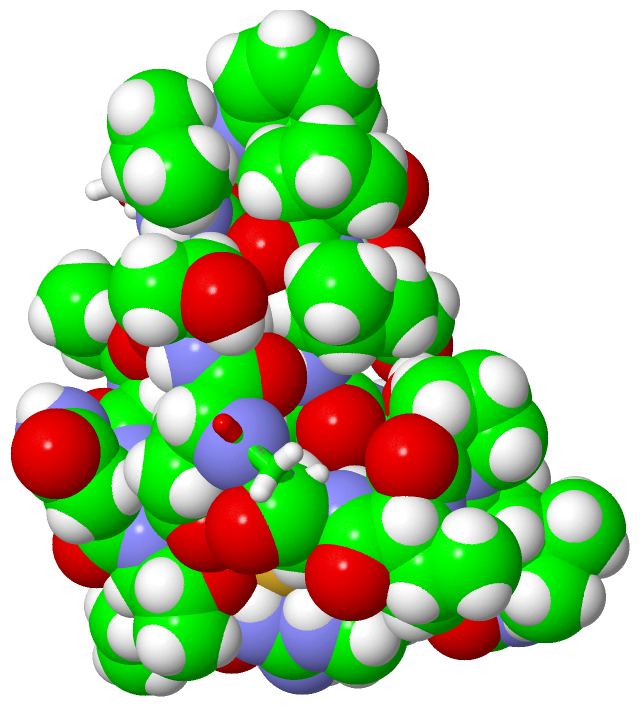
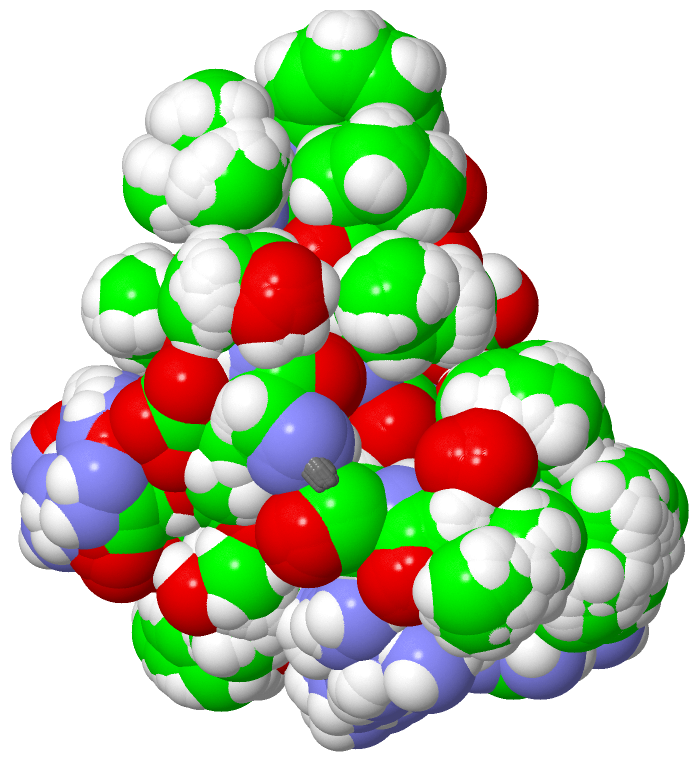
NMR Structure (24, 24)
No.NameEvidenceResiduesDescription
01AC1SOFTWAREGLY A:2 , DGL A:3 , GLY A:14 , DCY A:15 , DAR A:24 , DVA A:29BINDING SITE FOR RESIDUE DCY A 1
02AC2SOFTWAREDCY A:1 , GLY A:2 , DCY A:10 , DSG A:11 , DTH A:12 , DPR A:20 , DVA A:21BINDING SITE FOR RESIDUE DGL A 3
03AC3SOFTWAREGLY A:2 , DGL A:3 , DCY A:5 , DVA A:6 , GLY A:8 , DTH A:9 , DPR A:20BINDING SITE FOR RESIDUE DTH A 4
04AC4SOFTWAREDGL A:3 , DTH A:4 , DVA A:6 , GLY A:7 , GLY A:8 , DTH A:9 , DTH A:16 , DCY A:17 , DSN A:18BINDING SITE FOR RESIDUE DCY A 5
05AC5SOFTWAREDTH A:4 , DCY A:5 , GLY A:7 , GLY A:8 , DTR A:19BINDING SITE FOR RESIDUE DVA A 6
06AC6SOFTWAREDGL A:3 , DTH A:4 , DCY A:5 , DVA A:6 , GLY A:7 , GLY A:8 , DCY A:10BINDING SITE FOR RESIDUE DTH A 9
07AC7SOFTWAREDGL A:3 , DTH A:4 , DCY A:5 , GLY A:8 , DTH A:9 , DTH A:16BINDING SITE FOR RESIDUE DCY A 10
08AC8SOFTWAREDGL A:3 , DTH A:4 , DCY A:10 , DTH A:12 , DPR A:13BINDING SITE FOR RESIDUE DSG A 11
09AC9SOFTWAREDCY A:1 , DGL A:3 , DCY A:10 , DSG A:11 , DPR A:13BINDING SITE FOR RESIDUE DTH A 12
10BC1SOFTWAREDGL A:3 , DSG A:11 , DTH A:12 , GLY A:14BINDING SITE FOR RESIDUE DPR A 13
11BC2SOFTWAREDCY A:1 , DCY A:10 , DTH A:12 , GLY A:14 , DTH A:16 , DAR A:24BINDING SITE FOR RESIDUE DCY A 15
12BC3SOFTWAREDCY A:5 , GLY A:14BINDING SITE FOR RESIDUE DTH A 16
13BC4SOFTWAREDTH A:4 , DCY A:5 , DVA A:6 , GLY A:8 , DTH A:16 , DSN A:18BINDING SITE FOR RESIDUE DCY A 17
14BC5SOFTWAREDCY A:5 , DTH A:16 , DCY A:17 , DTH A:23 , GLY A:26BINDING SITE FOR RESIDUE DSN A 18
15BC6SOFTWAREDTH A:4 , DCY A:5 , DVA A:6 , DCY A:17 , DSN A:18 , DPR A:20BINDING SITE FOR RESIDUE DTR A 19
16BC7SOFTWAREDGL A:3 , DTH A:4 , DCY A:5 , DCY A:17 , DSN A:18 , DTR A:19 , DVA A:21BINDING SITE FOR RESIDUE DPR A 20
17BC8SOFTWAREGLY A:2 , DGL A:3 , DTH A:4 , DCY A:5 , DTH A:16 , DCY A:17 , DSN A:18 , DPR A:20BINDING SITE FOR RESIDUE DVA A 21
18BC9SOFTWAREDCY A:1 , GLY A:2 , DGL A:3 , DCY A:5 , DCY A:10 , DTH A:16 , DPR A:20 , DVA A:21BINDING SITE FOR RESIDUE DCY A 22
19CC1SOFTWAREDCY A:1 , DGL A:3 , DCY A:5 , GLY A:14 , DTH A:16 , DSN A:18 , DAR A:24 , GLY A:26BINDING SITE FOR RESIDUE DTH A 23
20CC2SOFTWAREDCY A:1 , DTH A:12 , GLY A:14 , DCY A:15 , DTH A:23 , DSG A:25BINDING SITE FOR RESIDUE DAR A 24
21CC3SOFTWAREGLY A:14 , DTH A:23 , DAR A:24BINDING SITE FOR RESIDUE DSG A 25
22CC4SOFTWAREDCY A:1 , DTH A:23 , DAR A:24 , DSG A:25 , DPR A:28 , DVA A:29BINDING SITE FOR RESIDUE DLE A 27
23CC5SOFTWAREDCY A:1 , GLY A:2 , DSN A:18 , DTH A:23 , DAR A:24BINDING SITE FOR RESIDUE DPR A 28
24CC6SOFTWAREDCY A:1 , DCY A:15 , DTH A:23 , DAR A:24 , DPR A:28BINDING SITE FOR RESIDUE DVA A 29

(-) SS Bonds  (3, 3)

NMR Structure
No.Residues
1A:1 -A:15
2A:5 -A:17
3A:10 -A:22

(-) Cis Peptide Bonds  (1, 20)

NMR Structure
No.ModelResidues
11, 2, 3, 4, 5, 6, 7, 8, 9, 10, 11, 12, 13, 14, 15, 16, 17, 18, 19, 20Dtr A:19 -Dpr A:20

 Sequence-Structure Mapping

(-) SAPs(SNPs)/Variants  (0, 0)

(no "SAP(SNP)/Variant" information available for 2JUE)

(-) PROSITE Motifs  (1, 1)

NMR Structure (1, 1)
 PROSITEUniProtKBPDB
No.IDACDescriptionIDLocationCountLocation
1CYCLOTIDE_MOEBIUSPS60009 Cyclotides Moebius subfamily signature.KAB1_OLDAF93-102  1A:2-8
NMR Structure * (1, 1)
 PROSITEUniProtKBPDB
No.IDACDescriptionIDLocationCountLocation
1CYCLOTIDE_MOEBIUSPS60009 Cyclotides Moebius subfamily signature.KAB1_OLDAF93-102  1A:2-8

(-) Exons   (0, 0)

(no "Exon" information available for 2JUE)

(-) Sequences/Alignments

NMR Structure
   Reformat: Number of residues per line =  ('0' or empty: single-line sequence representation)
  Number of residues per labelling interval =   
  UniProt sequence: complete  aligned part    
   Show mapping: SCOP domains CATH domains Pfam domains Secondary structure (by author)
SAPs(SNPs) PROSITE motifs Exons
(details for a mapped element are shown in a popup box when the mouse pointer rests over it)
Chain A from PDB  Type:PROTEIN  Length:29
 aligned with KAB1_OLDAF | P56254 from UniProtKB/Swiss-Prot  Length:124

    Alignment length:29
                                   102       112         
           KAB1_OLDAF    93 CGETCVGGTCNTPGCTCSWPVCTRNGLPS 121
               SCOP domains ----------------------------- SCOP domains
               CATH domains ----------------------------- CATH domains
               Pfam domains Cyclotide-2jueA01 A:1-25 ---- Pfam domains
         Sec.struct. author ..............eeee..eeee..... Sec.struct. author
                 SAPs(SNPs) ----------------------------- SAPs(SNPs)
                    PROSITE CYCLOTIDE_------------------- PROSITE
                 Transcript ----------------------------- Transcript
                 2jue A   1 xGxxxxGGxxxxxGxxxxxxxxxxxGxxx  29
                            | ||||  10||| ||||20||||| |||
                            | ||||  ||12-DTH||||22-DCY|||
                            1-DCY|  |||13-DPR||||23-DTH||
                              3-DGL |||  15-DCY|| 24-DAR|
                               4-DTH|||   16-DTH|  25-DSG
                                5-DCY||    17-DCY    27-DLE
                                 6-DVA|     18-DSN    28-DPR
                                    9-DTH    19-DTR    29-DVA
                                    10-DCY    20-DPR     
                                     11-DSG    21-DVA    

   Legend:   → Mismatch (orange background)
  - → Gap (green background, '-', border residues have a numbering label)
    → Modified Residue (blue background, lower-case, 'x' indicates undefined single-letter code, labelled with number + name)
  x → Chemical Group (purple background, 'x', labelled with number + name, e.g. ACE or NH2)
  extra numbering lines below/above indicate numbering irregularities and modified residue names etc., number ends below/above '|'

 Classification and Annotation

(-) SCOP Domains  (0, 0)

(no "SCOP Domain" information available for 2JUE)

(-) CATH Domains  (0, 0)

(no "CATH Domain" information available for 2JUE)

(-) Pfam Domains  (1, 1)

NMR Structure

(-) Gene Ontology  (4, 4)

NMR Structure(hide GO term definitions)
Chain A   (KAB1_OLDAF | P56254)
biological process
    GO:0019835    cytolysis    The rupture of cell membranes and the loss of cytoplasm.
    GO:0006952    defense response    Reactions, triggered in response to the presence of a foreign body or the occurrence of an injury, which result in restriction of damage to the organism attacked or prevention/recovery from the infection caused by the attack.
    GO:0042742    defense response to bacterium    Reactions triggered in response to the presence of a bacterium that act to protect the cell or organism.
    GO:0044179    hemolysis in other organism    The cytolytic destruction of red blood cells, with the release of intracellular hemoglobin, in one organism by another.

 Visualization

(-) Interactive Views

NMR Structure
  Complete Structure
    Jena3D(integrated viewing of ligand, site, SAP, PROSITE, SCOP information)
    WebMol | AstexViewer[tm]@PDBe
(Java Applets, require no local installation except for Java; loading may be slow)
    STRAP
(Java WebStart application, automatic local installation, requires Java; full application with system access!)
    RasMol
(require local installation)
    Molscript (VRML)
(requires installation of a VRML viewer; select preferred view via VRML and generate a mono or stereo PDF format file)
 
  Ligands, Modified Residues, Ions
    DAR  [ RasMol | Jena3D ]  +environment [ RasMol | Jena3D ]
    DCY  [ RasMol | Jena3D ]  +environment [ RasMol | Jena3D ]
    DGL  [ RasMol | Jena3D ]  +environment [ RasMol | Jena3D ]
    DLE  [ RasMol | Jena3D ]  +environment [ RasMol | Jena3D ]
    DPR  [ RasMol | Jena3D ]  +environment [ RasMol | Jena3D ]
    DSG  [ RasMol | Jena3D ]  +environment [ RasMol | Jena3D ]
    DSN  [ RasMol | Jena3D ]  +environment [ RasMol | Jena3D ]
    DTH  [ RasMol | Jena3D ]  +environment [ RasMol | Jena3D ]
    DTR  [ RasMol | Jena3D ]  +environment [ RasMol | Jena3D ]
    DVA  [ RasMol | Jena3D ]  +environment [ RasMol | Jena3D ]
 
  Sites
    AC1  [ RasMol ]  +environment [ RasMol ]
    AC2  [ RasMol ]  +environment [ RasMol ]
    AC3  [ RasMol ]  +environment [ RasMol ]
    AC4  [ RasMol ]  +environment [ RasMol ]
    AC5  [ RasMol ]  +environment [ RasMol ]
    AC6  [ RasMol ]  +environment [ RasMol ]
    AC7  [ RasMol ]  +environment [ RasMol ]
    AC8  [ RasMol ]  +environment [ RasMol ]
    AC9  [ RasMol ]  +environment [ RasMol ]
    BC1  [ RasMol ]  +environment [ RasMol ]
    BC2  [ RasMol ]  +environment [ RasMol ]
    BC3  [ RasMol ]  +environment [ RasMol ]
    BC4  [ RasMol ]  +environment [ RasMol ]
    BC5  [ RasMol ]  +environment [ RasMol ]
    BC6  [ RasMol ]  +environment [ RasMol ]
    BC7  [ RasMol ]  +environment [ RasMol ]
    BC8  [ RasMol ]  +environment [ RasMol ]
    BC9  [ RasMol ]  +environment [ RasMol ]
    CC1  [ RasMol ]  +environment [ RasMol ]
    CC2  [ RasMol ]  +environment [ RasMol ]
    CC3  [ RasMol ]  +environment [ RasMol ]
    CC4  [ RasMol ]  +environment [ RasMol ]
    CC5  [ RasMol ]  +environment [ RasMol ]
    CC6  [ RasMol ]  +environment [ RasMol ]
 
  Cis Peptide Bonds
    Dtr A:19 - Dpr A:20   [ RasMol ]  
 

(-) Still Images

Jmol
  protein: cartoon or spacefill or dots and stick; nucleic acid: cartoon and stick; ligands: spacefill; active site: stick
Molscript
  protein, nucleic acid: cartoon; ligands: spacefill; active site: ball and stick

 Databases and Analysis Tools

(-) Databases

Access by PDB/NDB ID
  2jue
    Family and Domain InformationProDom | SYSTERS
    General Structural InformationGlycoscienceDB | MMDB | NDB | OCA | PDB | PDBe | PDBj | PDBsum | PDBWiki | PQS | PROTEOPEDIA
    Orientation in MembranesOPM
    Protein SurfaceSURFACE
    Secondary StructureDSSP (structure derived) | HSSP (homology derived)
    Structural GenomicsGeneCensus
    Structural NeighboursCE | VAST
    Structure ClassificationCATH | Dali | SCOP
    Validation and Original DataBMRB Data View | BMRB Restraints Grid | EDS | PROCHECK | RECOORD | WHAT_CHECK
 
Access by UniProt ID/Accession number
  KAB1_OLDAF | P56254
    Comparative Protein Structure ModelsModBase
    Genomic InformationEnsembl
    Protein-protein InteractionDIP
    Sequence, Family and Domain InformationInterPro | Pfam | SMART | UniProtKB/SwissProt
 
Access by Enzyme Classificator   (EC Number)
  (no 'Enzyme Classificator' available)
    General Enzyme InformationBRENDA | EC-PDB | Enzyme | IntEnz
    PathwayKEGG | MetaCyc
 
Access by Disease Identifier   (MIM ID)
  (no 'MIM ID' available)
    Disease InformationOMIM
 
Access by GenAge ID
  (no 'GenAge ID' available)
    Age Related InformationGenAge

(-) Analysis Tools

Access by PDB/NDB ID
    Domain InformationXDom
    Interatomic Contacts of Structural UnitsCSU
    Ligand-protein ContactsLPC
    Protein CavitiescastP
    Sequence and Secondary StructurePDBCartoon
    Structure AlignmentSTRAP(Java WebStart application, automatic local installation, requires Java; full application with system access!)
    Structure and Sequence BrowserSTING
 
Access by UniProt ID/Accession number
  KAB1_OLDAF | P56254
    Protein Disorder PredictionDisEMBL | FoldIndex | GLOBPLOT (for more information see DisProt)

 Related Entries

(-) Entries Sharing at Least One Protein Chain (UniProt ID)

UniProtKB/Swiss-Prot
        KAB1_OLDAF | P562541jjz 1k48 1kal 1n1u 1nb1 1orx 1znu 2f2i 2f2j 2khb 2mh1 2mn1 4ttm 4ttn 4tto

(-) Related Entries Specified in the PDB File

1nb1 THIS ENTRY CONTAINS THE ALL-L VERSION OF KALATA B1