|
|
|
|
 Description
|
|
Compounds
|
||||||||||||||||||||
Chains, Units
Summary Information (see also Sequences/Alignments below) |
Ligands, Modified Residues, Ions (0, 0)| (no "Ligand,Modified Residues,Ions" information available for 1JJZ) |
Sites (0, 0)| (no "Site" information available for 1JJZ) |
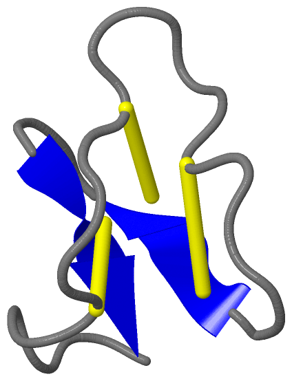
SS Bonds (3, 3)
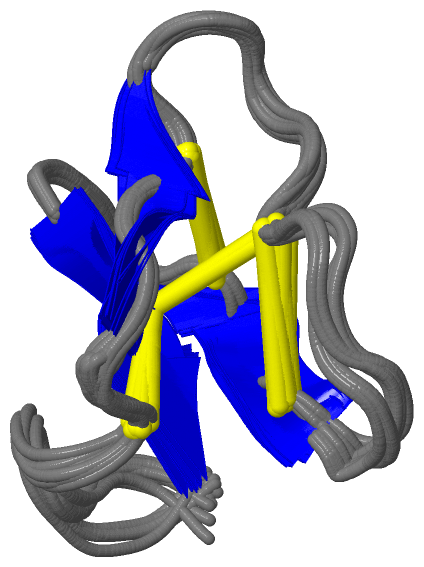
NMR Structure
|
||||||||||||||||
Cis Peptide Bonds (1, 20)
NMR Structure
|
||||||||||
SAPs(SNPs)/Variants (0, 0)| (no "SAP(SNP)/Variant" information available for 1JJZ) |
PROSITE Motifs (2, 2)
NMR Structure (2, 2)
|
||||||||||||||||||||||||||||||||
Exons (0, 0)| (no "Exon" information available for 1JJZ) |
Sequences/Alignments
NMR StructureChain A from PDB Type:PROTEIN Length:29 aligned with KAB1_OLDAF | P56254 from UniProtKB/Swiss-Prot Length:124 Alignment length:29 97 107 KAB1_OLDAF 88 KGLPVCGETCVGGTCNTPGCTCSWPVCTR 116 SCOP domains d1jjza_ A: Kalata B1 SCOP domains CATH domains ----------------------------- CATH domains Pfam domains -Cyclotide-1jjzA01 A:9-36 Pfam domains SAPs(SNPs) ----------------------------- SAPs(SNPs) PROSITE (1) -CYCLOTIDE PDB: A:9-36 PROSITE (1) PROSITE (2) -----CYCLOTIDE_-------------- PROSITE (2) Transcript ----------------------------- Transcript 1jjz A 8 NGLPVCGETCVGGTCNTPGCTCSWPVCTR 36 17 27
|
||||||||||||||||||||
SCOP Domains (1, 1)
NMR Structure
 |
CATH Domains (0, 0)| (no "CATH Domain" information available for 1JJZ) |
Pfam Domains (1, 1)
NMR Structure
|
Gene Ontology (4, 4)|
NMR Structure(hide GO term definitions) Chain A (KAB1_OLDAF | P56254)
|
||||||||||||||||||||||||||||||
Interactive Views
|
|||||||||||||||||||||||||||||||||||||||||||||||||||||||||||||||||||||||||||||||||||||||||||||||||||||||||||||||||||||
Still Images
|
||||||||||||||||
Databases
|
||||||||||||||||||||||||||||||||||||||||||||||||||||||||||||||||||||||||||||||||||||||||||||||||||||||||||||||||||||||||||||||||||||||||||||||||||||||||||||||||
Analysis Tools
|
|||||||||||||||||||||||||||||||||||||||||||||||||||||||||||||
Entries Sharing at Least One Protein Chain (UniProt ID)
Related Entries Specified in the PDB File
|
|