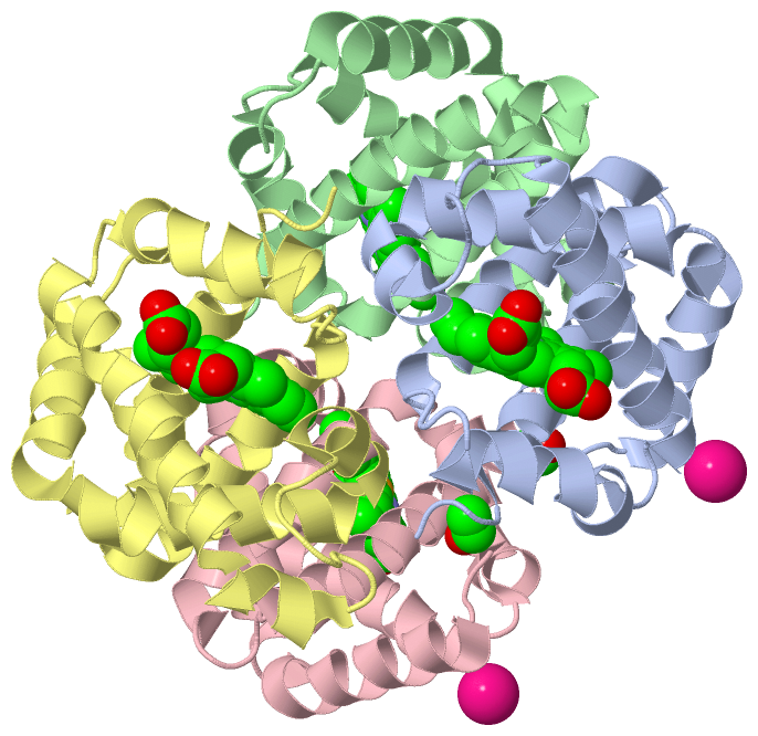
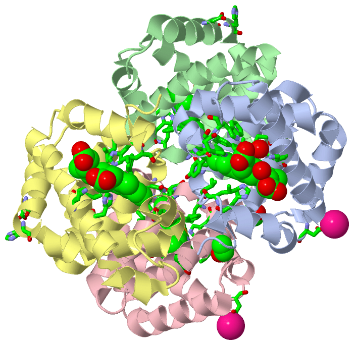
Show PDB file:   
         Plain Text   HTML   (compressed file size)
QuickSearch:   
by PDB,NDB,UniProt,PROSITE Code or Search Term(s)  
(-)Asym./Biol. Unit - manually
(-)Asym./Biol. Unit
(-)Asym./Biol. Unit - sites
collapse expand < >
Image Asym./Biol. Unit - manually
Asym./Biol. Unit - manually  (Jmol Viewer)
Image Asym./Biol. Unit
Asym./Biol. Unit  (Jmol Viewer)
Image Asym./Biol. Unit - sites
Asym./Biol. Unit - sites  (Jmol Viewer)

(-) Description

Title :  CRYSTAL STRUCTURE OF DEOXY HEMOGLOBIN FROM TREMATOMUS BERNACCHII AT PH 8.4
 
Authors :  L. Mazzarella, A. Vergara, L. Vitagliano, A. Merlino, G. Bonomi, S. Scala, C. Verde, G. Di Prisco
Date :  07 Jun 06  (Deposition) - 29 Aug 06  (Release) - 24 Feb 09  (Revision)
Method :  X-RAY DIFFRACTION
Resolution :  1.78
Chains :  Asym./Biol. Unit :  A,B,C,D
Keywords :  Root Effect, Ph, Cooperativity, Hemoglobin, Allostery, High Resolution, Transport Protein (Keyword Search: [Gene Ontology, PubMed, Web (Google))
 
Reference :  L. Mazzarella, A. Vergara, L. Vitagliano, A. Merlino, G. Bonomi, S. Scala, C. Verde, G. Di Prisco
High Resolution Crystal Structure Of Deoxy Hemoglobin From Trematomus Bernacchii At Different Ph Values: The Role Of Histidine Residues In Modulating The Strength Of The Root Effect.
Proteins V. 65 490 2006
PubMed-ID: 16909420  |  Reference-DOI: 10.1002/PROT.21114
(for further references see the PDB file header)

(-) Compounds

Molecule 1 - HEMOGLOBIN ALPHA SUBUNIT
    ChainsA, C
    Organism CommonEMERALD ROCKCOD
    Organism ScientificTREMATOMUS BERNACCHII
    Organism Taxid40690
    SynonymHEMOGLOBIN ALPHA CHAIN, ALPHA-GLOBIN
 
Molecule 2 - HEMOGLOBIN BETA SUBUNIT
    ChainsB, D
    Organism CommonEMERALD ROCKCOD
    Organism ScientificTREMATOMUS BERNACCHII
    Organism Taxid40690
    SynonymHEMOGLOBIN BETA CHAIN, BETA-GLOBIN

 Structural Features

(-) Chains, Units

  1234
Asymmetric/Biological Unit ABCD

Summary Information (see also Sequences/Alignments below)

(-) Ligands, Modified Residues, Ions  (3, 8)

Asymmetric/Biological Unit (3, 8)
No.NameCountTypeFull Name
1ACE2Mod. Amino AcidACETYL GROUP
2HEM4Ligand/IonPROTOPORPHYRIN IX CONTAINING FE
3K2Ligand/IonPOTASSIUM ION

(-) Sites  (6, 6)

Asymmetric Unit (6, 6)
No.NameEvidenceResiduesDescription
1AC1SOFTWAREASP B:13 , HIS B:17 , ASP C:75 , HOH C:1375 , HOH C:1376 , HOH C:1380BINDING SITE FOR RESIDUE K C 1001
2AC2SOFTWAREASP A:75 , HOH A:1378 , HOH A:1379 , ASP D:13 , HIS D:17BINDING SITE FOR RESIDUE K A 1002
3AC3SOFTWARETYR A:42 , PHE A:43 , HIS A:45 , TRP A:46 , HIS A:59 , LYS A:62 , VAL A:63 , HIS A:88 , LEU A:92 , VAL A:94 , ASN A:98 , LEU A:102 , HOH A:1061 , HOH A:1361BINDING SITE FOR RESIDUE HEM A 200
4AC4SOFTWARETHR B:38 , HIS B:41 , PHE B:42 , HIS B:63 , VAL B:67 , LEU B:91 , HIS B:92 , LEU B:96 , PHE B:103BINDING SITE FOR RESIDUE HEM B 400
5AC5SOFTWARETYR C:42 , PHE C:43 , HIS C:45 , TRP C:46 , HIS C:59 , LYS C:62 , VAL C:63 , LEU C:84 , HIS C:88 , LEU C:92 , ASN C:98 , PHE C:99 , LEU C:102 , HOH C:1264 , HOH C:1328BINDING SITE FOR RESIDUE HEM C 600
6AC6SOFTWARETHR D:38 , HIS D:63 , LYS D:66 , VAL D:67 , HIS D:92 , LEU D:96 , ASN D:102 , PHE D:103 , LEU D:141BINDING SITE FOR RESIDUE HEM D 800

(-) SS Bonds  (0, 0)

(no "SS Bond" information available for 2H8D)

(-) Cis Peptide Bonds  (0, 0)

(no "Cis Peptide Bond" information available for 2H8D)

 Sequence-Structure Mapping

(-) SAPs(SNPs)/Variants  (0, 0)

(no "SAP(SNP)/Variant" information available for 2H8D)

(-) PROSITE Motifs  (1, 4)

Asymmetric/Biological Unit (1, 4)
 PROSITEUniProtKBPDB
No.IDACDescriptionIDLocationCountLocation
1GLOBINPS01033 Globin family profile.HBA_TREBE2-142
 
  2A:2-142
C:2-142
HBB_TREBE5-147
 
  2B:4-146
D:4-146

(-) Exons   (0, 0)

(no "Exon" information available for 2H8D)

(-) Sequences/Alignments

Asymmetric/Biological Unit
   Reformat: Number of residues per line =  ('0' or empty: single-line sequence representation)
  Number of residues per labelling interval =   
  UniProt sequence: complete  aligned part    
   Show mapping: SCOP domains CATH domains Pfam domains Secondary structure (by author)
SAPs(SNPs) PROSITE motifs Exons
(details for a mapped element are shown in a popup box when the mouse pointer rests over it)
Chain A from PDB  Type:PROTEIN  Length:143
 aligned with HBA_TREBE | P80043 from UniProtKB/Swiss-Prot  Length:142

    Alignment length:143
                             1                                                                                                                                             
                             |       9        19        29        39        49        59        69        79        89        99       109       119       129       139   
            HBA_TREBE     - -SLSDKDKAAVRALWSKIGKSADAIGNDALSRMIVVYPQTKTYFSHWPDVTPGSPHIKAHGKKVMGGIALAVSKIDDLKTGLMELSEQHAYKLRVDPANFKILNHCILVVISTMFPKEFTPEAHVSLDKFLSGVALALAERYR 142
               SCOP domains d2h8da_ A: Hemoglobin, alpha-chain                                                                                                              SCOP domains
               CATH domains -2h8dA00 A:1-142 Globins                                                                                                                        CATH domains
               Pfam domains ----------------------------------------------------------------------------------------------------------------------------------------------- Pfam domains
         Sec.struct. author ...hhhhhhhhhhhhhhhh.hhhhhhhhhhhhhhhhhhhhhhhhh........hhhhhhhhhhhhhhhhhhhhh..hhhhhhhhhhhhhhh.....hhhhhhhhhhhhhhhhhhh....hhhhhhhhhhhhhhhhhhhh.... Sec.struct. author
                 SAPs(SNPs) ----------------------------------------------------------------------------------------------------------------------------------------------- SAPs(SNPs)
                    PROSITE --GLOBIN  PDB: A:2-142 UniProt: 2-142                                                                                                           PROSITE
                 Transcript ----------------------------------------------------------------------------------------------------------------------------------------------- Transcript
                 2h8d A   0 xSLSDKDKAAVRALWSKIGKSADAIGNDALSRMIVVYPQTKTYFSHWPDVTPGSPHIKAHGKKVMGGIALAVSKIDDLKTGLMELSEQHAYKLRVDPANFKILNHCILVVISTMFPKEFTPEAHVSLDKFLSGVALALAERYR 142
                            |        9        19        29        39        49        59        69        79        89        99       109       119       129       139   
                            |                                                                                                                                              
                            0-ACE                                                                                                                                          

Chain B from PDB  Type:PROTEIN  Length:146
 aligned with HBB_TREBE | P80044 from UniProtKB/Swiss-Prot  Length:147

    Alignment length:146
                                    11        21        31        41        51        61        71        81        91       101       111       121       131       141      
            HBB_TREBE     2 VEWTDKERSIISDIFSHMDYDDIGPKALSRCLIVYPWTQRHFSGFGNLYNAEAIIGNANVAAHGIKVLHGLDRGVKNMDNIAATYADLSTLHSEKLHVDPDNFKLLSDCITIVLAAKMGHAFTAETQGAFQKFLAVVVSALGKQYH 147
               SCOP domains d2h8db_ B: Hemoglobin, beta-chain                                                                                                                  SCOP domains
               CATH domains 2h8dB00 B:1-146 Globins                                                                                                                            CATH domains
               Pfam domains -------------------------------------------------------------------------------------------------------------------------------------------------- Pfam domains
         Sec.struct. author ...hhhhhhhhhhhhhhhhhhhhhhhhhhhhhhhhhhhhhhhhhhh...hhhhhhhhhhhhhhhhhhhhhhhhhhh...hhhhhhhhhhhhhhhh...hhhhhhhhhhhhhhhhhhhhhhhhhhhhhhhhhhhhhhhhhhhhh... Sec.struct. author
                 SAPs(SNPs) -------------------------------------------------------------------------------------------------------------------------------------------------- SAPs(SNPs)
                PROSITE (2) ---GLOBIN  PDB: B:4-146 UniProt: 5-147                                                                                                             PROSITE (2)
                 Transcript -------------------------------------------------------------------------------------------------------------------------------------------------- Transcript
                 2h8d B   1 VEWTDKERSIISDIFSHMDYDDIGPKALSRCLIVYPWTQRHFSGFGNLYNAEAIIGNANVAAHGIKVLHGLDRGVKNMDNIAATYADLSTLHSEKLHVDPDNFKLLSDCITIVLAAKMGHAFTAETQGAFQKFLAVVVSALGKQYH 146
                                    10        20        30        40        50        60        70        80        90       100       110       120       130       140      

Chain C from PDB  Type:PROTEIN  Length:143
 aligned with HBA_TREBE | P80043 from UniProtKB/Swiss-Prot  Length:142

    Alignment length:143
                             1                                                                                                                                             
                             |       9        19        29        39        49        59        69        79        89        99       109       119       129       139   
            HBA_TREBE     - -SLSDKDKAAVRALWSKIGKSADAIGNDALSRMIVVYPQTKTYFSHWPDVTPGSPHIKAHGKKVMGGIALAVSKIDDLKTGLMELSEQHAYKLRVDPANFKILNHCILVVISTMFPKEFTPEAHVSLDKFLSGVALALAERYR 142
               SCOP domains d2h8dc_ C: Hemoglobin, alpha-chain                                                                                                              SCOP domains
               CATH domains -2h8dC00 C:1-142 Globins                                                                                                                        CATH domains
               Pfam domains ----------------------------------------------------------------------------------------------------------------------------------------------- Pfam domains
         Sec.struct. author ...hhhhhhhhhhhhhhhh.hhhhhhhhhhhhhhhhhhhhhhhhh........hhhhhhhhhhhhhhhhhhhhh..hhhhhhhhhhhhhhhh....hhhhhhhhhhhhhhhhhhh....hhhhhhhhhhhhhhhhhhhhhhhh Sec.struct. author
                 SAPs(SNPs) ----------------------------------------------------------------------------------------------------------------------------------------------- SAPs(SNPs)
                    PROSITE --GLOBIN  PDB: C:2-142 UniProt: 2-142                                                                                                           PROSITE
                 Transcript ----------------------------------------------------------------------------------------------------------------------------------------------- Transcript
                 2h8d C   0 xSLSDKDKAAVRALWSKIGKSADAIGNDALSRMIVVYPQTKTYFSHWPDVTPGSPHIKAHGKKVMGGIALAVSKIDDLKTGLMELSEQHAYKLRVDPANFKILNHCILVVISTMFPKEFTPEAHVSLDKFLSGVALALAERYR 142
                            |        9        19        29        39        49        59        69        79        89        99       109       119       129       139   
                            0-ACE                                                                                                                                          

Chain D from PDB  Type:PROTEIN  Length:146
 aligned with HBB_TREBE | P80044 from UniProtKB/Swiss-Prot  Length:147

    Alignment length:146
                                    11        21        31        41        51        61        71        81        91       101       111       121       131       141      
            HBB_TREBE     2 VEWTDKERSIISDIFSHMDYDDIGPKALSRCLIVYPWTQRHFSGFGNLYNAEAIIGNANVAAHGIKVLHGLDRGVKNMDNIAATYADLSTLHSEKLHVDPDNFKLLSDCITIVLAAKMGHAFTAETQGAFQKFLAVVVSALGKQYH 147
               SCOP domains d2h8dd_ D: Hemoglobin, beta-chain                                                                                                                  SCOP domains
               CATH domains 2h8dD00 D:1-146 Globins                                                                                                                            CATH domains
               Pfam domains -------------------------------------------------------------------------------------------------------------------------------------------------- Pfam domains
         Sec.struct. author ...hhhhhhhhhhhhhhhhhhhhhhhhhhhhhhhhhhhhhhh.......hhhhhhhhhhhhhhhhhhhhhhhhhhhh..hhhhhhhhhhhhhhhh....hhhhhhhhhhhhhhhhhhhhhhhhhhhhhhhhhhhhhhhhhhh.... Sec.struct. author
                 SAPs(SNPs) -------------------------------------------------------------------------------------------------------------------------------------------------- SAPs(SNPs)
                PROSITE (2) ---GLOBIN  PDB: D:4-146 UniProt: 5-147                                                                                                             PROSITE (2)
                 Transcript -------------------------------------------------------------------------------------------------------------------------------------------------- Transcript
                 2h8d D   1 VEWTDKERSIISDIFSHMDYDDIGPKALSRCLIVYPWTQRHFSGFGNLYNAEAIIGNANVAAHGIKVLHGLDRGVKNMDNIAATYADLSTLHSEKLHVDPDNFKLLSDCITIVLAAKMGHAFTAETQGAFQKFLAVVVSALGKQYH 146
                                    10        20        30        40        50        60        70        80        90       100       110       120       130       140      

   Legend:   → Mismatch (orange background)
  - → Gap (green background, '-', border residues have a numbering label)
    → Modified Residue (blue background, lower-case, 'x' indicates undefined single-letter code, labelled with number + name)
  x → Chemical Group (purple background, 'x', labelled with number + name, e.g. ACE or NH2)
  extra numbering lines below/above indicate numbering irregularities and modified residue names etc., number ends below/above '|'

 Classification and Annotation

(-) SCOP Domains  (2, 4)

Asymmetric/Biological Unit

(-) CATH Domains  (1, 4)

Asymmetric/Biological Unit

(-) Pfam Domains  (0, 0)

(no "Pfam Domain" information available for 2H8D)

(-) Gene Ontology  (8, 16)

Asymmetric/Biological Unit(hide GO term definitions)
Chain A,C   (HBA_TREBE | P80043)
molecular function
    GO:0020037    heme binding    Interacting selectively and non-covalently with heme, any compound of iron complexed in a porphyrin (tetrapyrrole) ring.
    GO:0005506    iron ion binding    Interacting selectively and non-covalently with iron (Fe) ions.
    GO:0046872    metal ion binding    Interacting selectively and non-covalently with any metal ion.
    GO:0019825    oxygen binding    Interacting selectively and non-covalently with oxygen (O2).
    GO:0005344    oxygen transporter activity    Enables the directed movement of oxygen into, out of or within a cell, or between cells.
biological process
    GO:0015671    oxygen transport    The directed movement of oxygen (O2) into, out of or within a cell, or between cells, by means of some agent such as a transporter or pore.
    GO:0006810    transport    The directed movement of substances (such as macromolecules, small molecules, ions) or cellular components (such as complexes and organelles) into, out of or within a cell, or between cells, or within a multicellular organism by means of some agent such as a transporter, pore or motor protein.
cellular component
    GO:0005833    hemoglobin complex    An iron-containing, oxygen carrying complex. In vertebrates it is made up of two pairs of associated globin polypeptide chains, each chain carrying a noncovalently bound heme prosthetic group.

Chain B,D   (HBB_TREBE | P80044)
molecular function
    GO:0020037    heme binding    Interacting selectively and non-covalently with heme, any compound of iron complexed in a porphyrin (tetrapyrrole) ring.
    GO:0005506    iron ion binding    Interacting selectively and non-covalently with iron (Fe) ions.
    GO:0046872    metal ion binding    Interacting selectively and non-covalently with any metal ion.
    GO:0019825    oxygen binding    Interacting selectively and non-covalently with oxygen (O2).
    GO:0005344    oxygen transporter activity    Enables the directed movement of oxygen into, out of or within a cell, or between cells.
biological process
    GO:0015671    oxygen transport    The directed movement of oxygen (O2) into, out of or within a cell, or between cells, by means of some agent such as a transporter or pore.
    GO:0006810    transport    The directed movement of substances (such as macromolecules, small molecules, ions) or cellular components (such as complexes and organelles) into, out of or within a cell, or between cells, or within a multicellular organism by means of some agent such as a transporter, pore or motor protein.
cellular component
    GO:0005833    hemoglobin complex    An iron-containing, oxygen carrying complex. In vertebrates it is made up of two pairs of associated globin polypeptide chains, each chain carrying a noncovalently bound heme prosthetic group.

 Visualization

(-) Interactive Views

Asymmetric/Biological Unit
  Complete Structure
    Jena3D(integrated viewing of ligand, site, SAP, PROSITE, SCOP information)
    WebMol | AstexViewer[tm]@PDBe
(Java Applets, require no local installation except for Java; loading may be slow)
    STRAP
(Java WebStart application, automatic local installation, requires Java; full application with system access!)
    RasMol
(require local installation)
    Molscript (VRML)
(requires installation of a VRML viewer; select preferred view via VRML and generate a mono or stereo PDF format file)
 
  Ligands, Modified Residues, Ions
    ACE  [ RasMol | Jena3D ]  +environment [ RasMol | Jena3D ]
    HEM  [ RasMol | Jena3D ]  +environment [ RasMol | Jena3D ]
    K  [ RasMol | Jena3D ]  +environment [ RasMol | Jena3D ]
 
  Sites
    AC1  [ RasMol ]  +environment [ RasMol ]
    AC2  [ RasMol ]  +environment [ RasMol ]
    AC3  [ RasMol ]  +environment [ RasMol ]
    AC4  [ RasMol ]  +environment [ RasMol ]
    AC5  [ RasMol ]  +environment [ RasMol ]
    AC6  [ RasMol ]  +environment [ RasMol ]
 
  Cis Peptide Bonds
(no "Cis Peptide Bonds" information available for 2h8d)
 

(-) Still Images

Jmol
  protein: cartoon or spacefill or dots and stick; nucleic acid: cartoon and stick; ligands: spacefill; active site: stick
Molscript
  protein, nucleic acid: cartoon; ligands: spacefill; active site: ball and stick

 Databases and Analysis Tools

(-) Databases

Access by PDB/NDB ID
  2h8d
    Family and Domain InformationProDom | SYSTERS
    General Structural InformationGlycoscienceDB | MMDB | NDB | OCA | PDB | PDBe | PDBj | PDBsum | PDBWiki | PQS | PROTEOPEDIA
    Orientation in MembranesOPM
    Protein SurfaceSURFACE
    Secondary StructureDSSP (structure derived) | HSSP (homology derived)
    Structural GenomicsGeneCensus
    Structural NeighboursCE | VAST
    Structure ClassificationCATH | Dali | SCOP
    Validation and Original DataBMRB Data View | BMRB Restraints Grid | EDS | PROCHECK | RECOORD | WHAT_CHECK
 
Access by UniProt ID/Accession number
  HBA_TREBE | P80043
    Comparative Protein Structure ModelsModBase
    Genomic InformationEnsembl
    Protein-protein InteractionDIP
    Sequence, Family and Domain InformationInterPro | Pfam | SMART | UniProtKB/SwissProt
  HBB_TREBE | P80044
    Comparative Protein Structure ModelsModBase
    Genomic InformationEnsembl
    Protein-protein InteractionDIP
    Sequence, Family and Domain InformationInterPro | Pfam | SMART | UniProtKB/SwissProt
 
Access by Enzyme Classificator   (EC Number)
  (no 'Enzyme Classificator' available)
    General Enzyme InformationBRENDA | EC-PDB | Enzyme | IntEnz
    PathwayKEGG | MetaCyc
 
Access by Disease Identifier   (MIM ID)
  (no 'MIM ID' available)
    Disease InformationOMIM
 
Access by GenAge ID
  (no 'GenAge ID' available)
    Age Related InformationGenAge

(-) Analysis Tools

Access by PDB/NDB ID
    Domain InformationXDom
    Interatomic Contacts of Structural UnitsCSU
    Ligand-protein ContactsLPC
    Protein CavitiescastP
    Sequence and Secondary StructurePDBCartoon
    Structure AlignmentSTRAP(Java WebStart application, automatic local installation, requires Java; full application with system access!)
    Structure and Sequence BrowserSTING
 
Access by UniProt ID/Accession number
  HBA_TREBE | P80043
    Protein Disorder PredictionDisEMBL | FoldIndex | GLOBPLOT (for more information see DisProt)
  HBB_TREBE | P80044
    Protein Disorder PredictionDisEMBL | FoldIndex | GLOBPLOT (for more information see DisProt)

 Related Entries

(-) Entries Sharing at Least One Protein Chain (UniProt ID)

UniProtKB/Swiss-Prot
        HBA_TREBE | P800431hbh 1pbx 1s5x 1s5y 2h8f 2peg 3gkv 3gqg 4g51 4iro 4odc
        HBB_TREBE | P800441hbh 1pbx 1s5x 1s5y 2h8f 2peg 3gkv 3gqg 4g51 4iro 4odc

(-) Related Entries Specified in the PDB File

2aa1 CRYSTAL STRUCTURE OF THE CATHODIC HEMOLGLOBIN ISOLATED FROM THE ANTARCTIC FISH TREMATOMUS NEWNESI
2h8f CRYSTAL STRUCTURE OF DEOXY HEMOGLOBIN FROM TREMATOMUS BERNACCHII AT PH 6.2