|
|
|
|
 Description
|
|
Compounds
|
||||||||||||||||||||||||||||||||||||||||||||||||
Chains, Units
Summary Information (see also Sequences/Alignments below) |
Ligands, Modified Residues, Ions (1, 13)
Asymmetric Unit (1, 13)
|
Sites (13, 13)
Asymmetric Unit (13, 13)
|
SS Bonds (0, 0)| (no "SS Bond" information available for 2FG4) |
Cis Peptide Bonds (0, 0)| (no "Cis Peptide Bond" information available for 2FG4) |
SAPs(SNPs)/Variants (2, 2)
Asymmetric Unit (2, 2)
|
||||||||||||||||||||||||||||||||||||||||||||||||||||||||||||||||||||||||||||||||||||||||||||||||||||||||||||||||||||||||||||||||||||
PROSITE Motifs (3, 3)
Asymmetric Unit (3, 3)
|
||||||||||||||||||||||||||||||||||||||||||||||||||||||||||||||||||||||||||||||||
Exons (4, 4)
Asymmetric Unit (4, 4)
|
||||||||||||||||||||||||||||||||||||||||||||||||||||||||||||||||||||||||
Sequences/Alignments
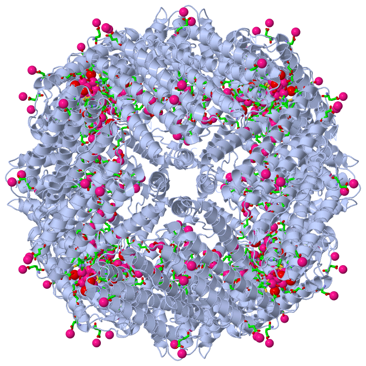
Asymmetric UnitChain A from PDB Type:PROTEIN Length:171 aligned with FRIL_HUMAN | P02792 from UniProtKB/Swiss-Prot Length:175 Alignment length:171 11 21 31 41 51 61 71 81 91 101 111 121 131 141 151 161 171 FRIL_HUMAN 2 SSQIRQNYSTDVEAAVNSLVNLYLQASYTYLSLGFYFDRDDVALEGVSHFFRELAEEKREGYERLLKMQNQRGGRALFQDIKKPAEDEWGKTPDAMKAAMALEKKLNQALLDLHALGSARTDPHLCDFLETHFLDEEVKLIKKMGDHLTNLHRLGGPEAGLGEYLFERLTL 172 SCOP domains d2fg4a_ A: (Apo)ferritin SCOP domains CATH domains 2fg4A00 A:5-175 [code=1.20.1260.10, no name defined] CATH domains Pfam domains --------------------------------------------------------------------------------------------------------------------------------------------------------------------------- Pfam domains SAPs(SNPs) ----------------------------I-----------------------------------------------------------------T---------------------------------------------------------------------------- SAPs(SNPs) PROSITE (1) -----FERRITIN_LIKE PDB: A:10-159 UniProt: 7-156 ---------------- PROSITE (1) PROSITE (2) --------------------------------------------------------FERRITIN_1 ----------------------------------------------FERRITIN_2 ----------------------------- PROSITE (2) Transcript 1 Exon 1.1 PDB: A:5-37 Exon 1.2 PDB: A:38-86 UniProt: 35-83 Exon 1.3 PDB: A:87-128 UniProt: 84-125 Exon 1.4 PDB: A:129-175 UniProt: 126-175 Transcript 1 2fg4 A 5 SSQIRQNYSTDVEAAVNSLVNLYLQASYTYLSLGFYFDRDDVALEGVSHFFRELAEEKREGYERLLKMQNQRGGRALFQDIKKPAEDEWGKTPDAMKAAMALEKKLNQALLDLHALGSARTDPHLCDFLETHFLDEEVKLIKKMGDHLTNLHRLGGPEAGLGEYLFERLTL 175 14 24 34 44 54 64 74 84 94 104 114 124 134 144 154 164 174
|
||||||||||||||||||||
SCOP Domains (1, 1)
Asymmetric Unit
 |
CATH Domains (1, 1)
Asymmetric Unit
|
Pfam Domains (0, 0)| (no "Pfam Domain" information available for 2FG4) |
Gene Ontology (12, 12)|
Asymmetric Unit(hide GO term definitions) Chain A (FRIL_HUMAN | P02792)
|
||||||||||||||||||||||||||||||||||||||||||||||||||||||||||||||||||||||||||||||||||||||||||
Interactive Views
|
||||||||||||||||||||||||||||||||||||||||||||||||||||||||||||||||||||||||||||||||||||||||||||||||||||||||||||||||||||||||||||||||||||||||||||||||||||||||||||||||||||||||||||||||||||||||||||||||||||||||||||||||||||||||||||
Still Images
|
||||||||||||||||
Databases
|
|||||||||||||||||||||||||||||||||||||||||||||||||||||||||||||||||||||||||||||||||||||||||||||||||||||||||||||||||||||||||||||||||||||||||||||||||||||||||||||||||||||||||||
Analysis Tools
|
|||||||||||||||||||||||||||||||||||||||||||||||||||||||||||||
Entries Sharing at Least One Protein Chain (UniProt ID)
Related Entries Specified in the PDB File
|
|