|
|
|
|
 Description
|
|
Compounds
|
||||||||||||||||||||||||||||||||||||||||||||||||
Chains, Units
Summary Information (see also Sequences/Alignments below) |
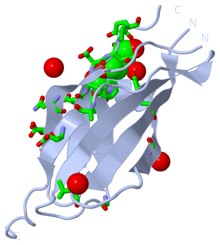
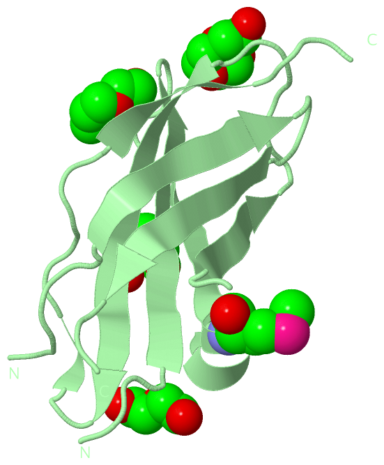
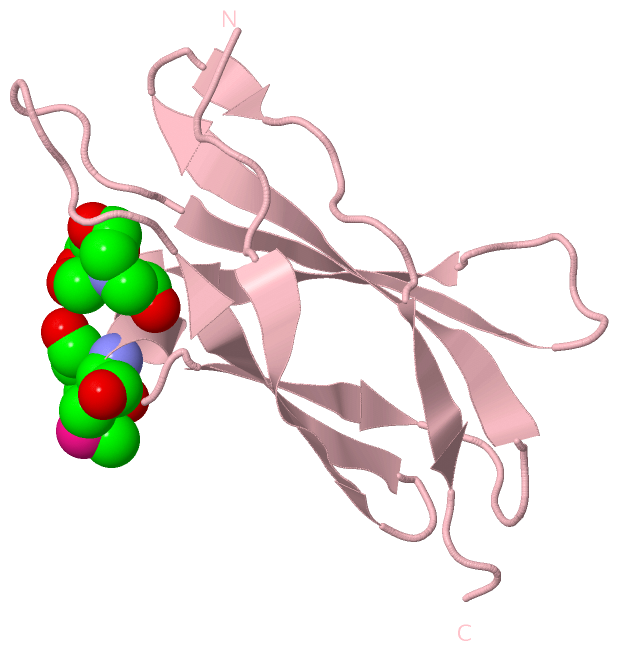
Ligands, Modified Residues, Ions (5, 11)
Asymmetric Unit (5, 11)
|
Sites (8, 8)
Asymmetric Unit (8, 8)
|
SS Bonds (0, 0)| (no "SS Bond" information available for 2E3V) |
Cis Peptide Bonds (0, 0)| (no "Cis Peptide Bond" information available for 2E3V) |
SAPs(SNPs)/Variants (0, 0)| (no "SAP(SNP)/Variant" information available for 2E3V) |
PROSITE Motifs (1, 4)
Asymmetric Unit (1, 4)
|
||||||||||||||||||||||||||||||||||||||||||||||||||||||||||||||||||||||||||||||||||||||||||||||||
Exons (3, 7)
Sequences/Alignments
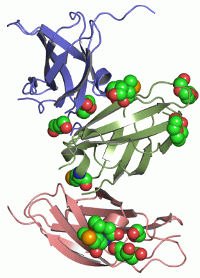
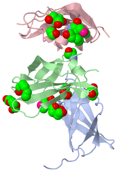
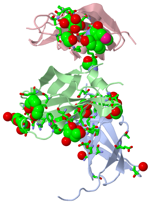
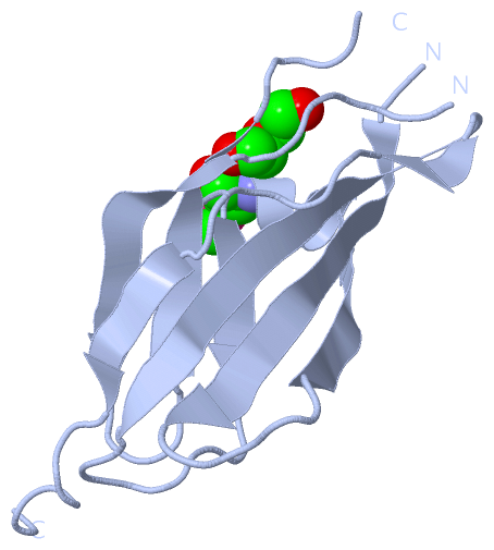
Asymmetric UnitChain A from PDB Type:PROTEIN Length:103 aligned with NCAM1_HUMAN | P13591 from UniProtKB/Swiss-Prot Length:858 Alignment length:106 517 527 537 547 557 567 577 587 597 607 NCAM1_HUMAN 508 DTPSSPSIDQVEPYSSTAQVQFDEPEATGGVPILKYKAEWRAVGEEVWHSKWYDAKEASMEGIVTIVGLKPETTYAVRLAALNGKGLGEISAASEFKTQPVQGEPS 613 SCOP domains d2e3va_ A: Neural cell adhes ion molecule 1, NCAM SCOP domains CATH domains ---------------------------------------------------------------------------------------------------------- CATH domains Pfam domains ---------------------------------------------------------------------------------------------------------- Pfam domains SAPs(SNPs) ---------------------------------------------------------------------------------------------------------- SAPs(SNPs) PROSITE -FN3 PDB: A:10-108 UniProt: 509-608 --FN3 PROSITE Transcript 1 (1) Exon 1.19b PDB: A:8-64 (gaps) UniProt: 507-564 --------------------------------------------1.28c Transcript 1 (1) Transcript 1 (2) --------------------------------------------------------Exon 1.20b PDB: A:64-109 UniProt: 564-609 ---- Transcript 1 (2) 2e3v A 8 SGPSSPSIDQVEPYSSTAQVQFDEPEAT--VPILKYKAEWRAVGEEVWHSKWYDAKEASmEGIVTIVGLKPETTYAVRLAALNGKGLGEISAASEFKTQPVR-EPS 112 17 27 | -| 47 57 67 77 87 97 107 | | 35 38 67-MSE 109 | 110 Chain B from PDB Type:PROTEIN Length:97 aligned with NCAM1_HUMAN | P13591 from UniProtKB/Swiss-Prot Length:858 Alignment length:101 517 527 537 547 557 567 577 587 597 607 NCAM1_HUMAN 508 DTPSSPSIDQVEPYSSTAQVQFDEPEATGGVPILKYKAEWRAVGEEVWHSKWYDAKEASMEGIVTIVGLKPETTYAVRLAALNGKGLGEISAASEFKTQPV 608 SCOP domains d2e3vb_ B: Neural cell adh esion molecule 1, NCAM SCOP domains CATH domains ----------------------------------------------------------------------------------------------------- CATH domains Pfam domains ----------------------------------------------------------------------------------------------------- Pfam domains SAPs(SNPs) ----------------------------------------------------------------------------------------------------- SAPs(SNPs) PROSITE -FN3 PDB: B:10-108 UniProt: 509-608 PROSITE Transcript 1 (1) Exon 1.19b PDB: B:8-64 (gaps) UniProt: 507-564 -------------------------------------------- Transcript 1 (1) Transcript 1 (2) --------------------------------------------------------Exon 1.20b PDB: B:64-108 UniProt: 564-609 Transcript 1 (2) 2e3v B 8 SGPSSPSIDQVEPYSSTAQVQFDEPE----VPILKYKAEWRAVGEEVWHSKWYDAKEASmEGIVTIVGLKPETTYAVRLAALNGKGLGEISAASEFKTQPV 108 17 27 | -| 47 57 67 77 87 97 107 33 38 67-MSE Chain C from PDB Type:PROTEIN Length:101 aligned with NCAM1_HUMAN | P13591 from UniProtKB/Swiss-Prot Length:858 Alignment length:101 517 527 537 547 557 567 577 587 597 607 NCAM1_HUMAN 508 DTPSSPSIDQVEPYSSTAQVQFDEPEATGGVPILKYKAEWRAVGEEVWHSKWYDAKEASMEGIVTIVGLKPETTYAVRLAALNGKGLGEISAASEFKTQPV 608 SCOP domains d2e3vc_ C: Neural cell adhesion molecule 1, NCAM SCOP domains CATH domains ----------------------------------------------------------------------------------------------------- CATH domains Pfam domains ----------------------------------------------------------------------------------------------------- Pfam domains SAPs(SNPs) ----------------------------------------------------------------------------------------------------- SAPs(SNPs) PROSITE -FN3 PDB: C:10-108 UniProt: 509-608 PROSITE Transcript 1 (1) Exon 1.19b PDB: C:8-64 UniProt: 507-564 [INCOMPLETE] -------------------------------------------- Transcript 1 (1) Transcript 1 (2) --------------------------------------------------------Exon 1.20b PDB: C:64-108 UniProt: 564-609 Transcript 1 (2) 2e3v C 8 SGPSSPSIDQVEPYSSTAQVQFDEPEATGGVPILKYKAEWRAVGEEVWHSKWYDAKEASmEGIVTIVGLKPETTYAVRLAALNGKGLGEISAASEFKTQPV 108 17 27 37 47 57 67 77 87 97 107 67-MSE
|
||||||||||||||||||||
SCOP Domains (1, 3)
Asymmetric Unit
 |
CATH Domains (0, 0)| (no "CATH Domain" information available for 2E3V) |
Pfam Domains (0, 0)| (no "Pfam Domain" information available for 2E3V) |
Gene Ontology (22, 22)|
Asymmetric Unit(hide GO term definitions) Chain A,B,C (NCAM1_HUMAN | P13591)
|
||||||||||||||||||||||||||||||||||||||||||||||||||||||||||||||||||||||||||||||||||||||||||||||||||||||||||||||||||||||||||||||||||||||||||||||||||||||
Interactive Views
|
|||||||||||||||||||||||||||||||||||||||||||||||||||||||||||||||||||||||||||||||||||||||||||||||||||||||||||||||||||||||||||||||||||||||||||||||||||||||||||||||||||||||||||||||||||||||||||||||||||||||||||||||||||||||||||||||
Still Images
|
||||||||||||||||
Databases
|
||||||||||||||||||||||||||||||||||||||||||||||||||||||||||||||||||||||||||||||||||||||||||||||||||||||||||||||||||||||||||||||||||||||||||||||||||||||||||||||||
Analysis Tools
|
|||||||||||||||||||||||||||||||||||||||||||||||||||||||||||||
Entries Sharing at Least One Protein Chain (UniProt ID)
Related Entries Specified in the PDB File
|
|