Show PDB file:   
         Plain Text   HTML   (compressed file size)
QuickSearch:   
by PDB,NDB,UniProt,PROSITE Code or Search Term(s)  
(-)Asymmetric Unit
(-)Asym. Unit - sites
(-)Biological Unit 1
(-)Biol. Unit 1 - sites
(-)Biological Unit 2
collapse expand < >
Image Asymmetric Unit
Asymmetric Unit  (Jmol Viewer)
Image Asym. Unit - sites
Asym. Unit - sites  (Jmol Viewer)
Image Biological Unit 1
Biological Unit 1  (Jmol Viewer)
Image Biol. Unit 1 - sites
Biol. Unit 1 - sites  (Jmol Viewer)
Image Biological Unit 2
Biological Unit 2  (Jmol Viewer)

(-) Description

Title :  CRYSTAL STRUCTURE OF TYPE I CYTOCHROME C3 FROM DESULFOVIBRIO AFRICANUS
 
Authors :  M. Czjzek, L. Pieulle, X. Morelli, F. Guerlesquin, E. C. Hatchikian
Date :  27 Apr 05  (Deposition) - 19 May 05  (Release) - 24 Feb 09  (Revision)
Method :  X-RAY DIFFRACTION
Resolution :  1.68
Chains :  Asym. Unit :  A,B
Biol. Unit 1:  A  (1x)
Biol. Unit 2:  B  (1x)
Keywords :  Basic Cytochrome C3, Electron Transfer, Sulfate Reducing Bacteria, Sad, Heme, Iron, Electron Transport (Keyword Search: [Gene Ontology, PubMed, Web (Google))
 
Reference :  L. Pieulle, X. Morelli, P. Gallice, E. Lojou, P. Barbier, M. Czjzek, P. Bianco, F. Guerlesquin, E. C. Hatchikian
The Type I / Type Ii Cytochrome C(3) Complex: An Electron Transfer Link In The Hydrogen-Sulfate Reduction Pathway.
J. Mol. Biol. V. 354 73 2005
PubMed-ID: 16226767  |  Reference-DOI: 10.1016/J.JMB.2005.09.036

(-) Compounds

Molecule 1 - BASIC CYTOCHROME C3
    ChainsA, B
    Organism ScientificDESULFOVIBRIO AFRICANUS
    Organism Taxid873
    Other DetailsFOUR C-TYPE HEME-GROUPS COVALENTLY LINKED BY CYSTEINE RESIDUES
    SynonymCYTOCHROME C3

 Structural Features

(-) Chains, Units

  12
Asymmetric Unit AB
Biological Unit 1 (1x)A 
Biological Unit 2 (1x) B

Summary Information (see also Sequences/Alignments below)

(-) Ligands, Modified Residues, Ions  (2, 10)

Asymmetric Unit (2, 10)
No.NameCountTypeFull Name
1CA2Ligand/IonCALCIUM ION
2HEC8Ligand/IonHEME C
Biological Unit 1 (1, 4)
No.NameCountTypeFull Name
1CA-1Ligand/IonCALCIUM ION
2HEC4Ligand/IonHEME C
Biological Unit 2 (1, 4)
No.NameCountTypeFull Name
1CA-1Ligand/IonCALCIUM ION
2HEC4Ligand/IonHEME C

(-) Sites  (10, 10)

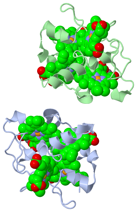
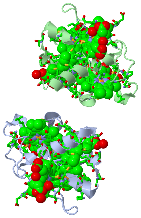
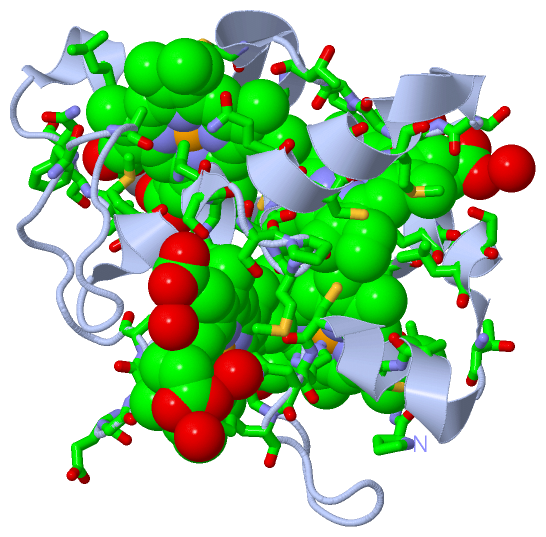
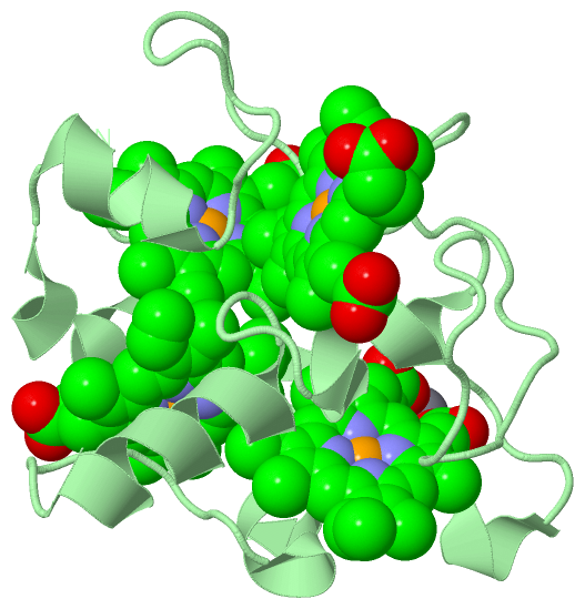
Asymmetric Unit (10, 10)
No.NameEvidenceResiduesDescription
01AC1SOFTWAREASP A:10 , LEU A:12 , GLU A:20 , TYR A:21 , HEC A:1118BINDING SITE FOR RESIDUE CA A1119
02AC2SOFTWAREASP B:10 , LEU B:12 , GLU B:20 , TYR B:21 , HEC B:1119BINDING SITE FOR RESIDUE CA B1120
03AC3SOFTWAREPRO A:1 , GLN A:2 , VAL A:3 , PRO A:4 , HIS A:11 , PHE A:25 , HIS A:27 , HIS A:30 , ALA A:39 , CYS A:40 , CYS A:43 , HIS A:44 , GLY A:54 , HEC A:1117 , HOH A:2130BINDING SITE FOR RESIDUE HEC A1115
04AC4SOFTWARECYS A:43 , HIS A:44 , HIS A:45 , LYS A:46 , CYS A:56 , GLU A:59 , GLY A:60 , CYS A:61 , HIS A:62 , ALA A:81 , SER A:84 , SER A:86 , MET A:88 , SER A:89 , HOH A:2131 , HOH A:2132 , HOH A:2133 , LYS B:28BINDING SITE FOR RESIDUE HEC A1116
05AC5SOFTWAREPHE A:25 , ALA A:29 , HIS A:30 , SER A:32 , LEU A:33 , THR A:35 , CYS A:43 , PRO A:87 , MET A:88 , SER A:89 , CYS A:90 , CYS A:93 , HIS A:94 , MET A:97 , THR A:103 , THR A:104 , GLY A:105 , PRO A:106 , HEC A:1115 , HOH A:2134BINDING SITE FOR RESIDUE HEC A1117
06AC6SOFTWAREILE A:9 , ASP A:10 , HIS A:11 , LEU A:12 , SER A:13 , ASN A:14 , LEU A:19 , GLU A:20 , TYR A:21 , VAL A:23 , PHE A:68 , ALA A:70 , MET A:79 , PHE A:82 , HIS A:83 , CYS A:90 , GLN A:91 , HIS A:94 , PRO A:106 , THR A:107 , CYS A:109 , CYS A:112 , HIS A:113 , CA A:1119BINDING SITE FOR RESIDUE HEC A1118
07AC7SOFTWAREGLN B:2 , HIS B:11 , PHE B:25 , HIS B:27 , HIS B:30 , ALA B:39 , CYS B:40 , CYS B:43 , HIS B:44 , ILE B:53 , GLY B:54 , GLY B:55 , HEC B:1118 , HOH B:2010 , HOH B:2081 , HOH B:2141 , HOH B:2142 , HOH B:2143 , HOH B:2144 , HOH B:2145BINDING SITE FOR RESIDUE HEC B1116
08AC8SOFTWARELYS A:28 , CYS B:43 , HIS B:45 , LYS B:46 , GLY B:55 , CYS B:56 , GLU B:59 , GLY B:60 , CYS B:61 , HIS B:62 , PRO B:75 , ALA B:81 , SER B:84 , SER B:86 , MET B:88 , SER B:89 , HOH B:2146 , HOH B:2147BINDING SITE FOR RESIDUE HEC B1117
09AC9SOFTWAREPHE B:25 , ALA B:29 , HIS B:30 , SER B:32 , LEU B:33 , THR B:35 , CYS B:43 , PRO B:87 , SER B:89 , CYS B:90 , CYS B:93 , HIS B:94 , MET B:97 , THR B:103 , THR B:104 , GLY B:105 , PRO B:106 , GLN B:115 , HEC B:1116 , HOH B:2148 , HOH B:2149BINDING SITE FOR RESIDUE HEC B1118
10BC1SOFTWAREILE B:9 , ASP B:10 , HIS B:11 , LEU B:12 , SER B:13 , ASN B:14 , LEU B:19 , GLU B:20 , TYR B:21 , VAL B:23 , PHE B:68 , ALA B:70 , MET B:79 , PHE B:82 , HIS B:83 , CYS B:90 , GLN B:91 , HIS B:94 , PRO B:106 , THR B:107 , CYS B:109 , CYS B:112 , HIS B:113 , CA B:1120BINDING SITE FOR RESIDUE HEC B1119

(-) SS Bonds  (0, 0)

(no "SS Bond" information available for 2BQ4)

(-) Cis Peptide Bonds  (0, 0)

(no "Cis Peptide Bond" information available for 2BQ4)

 Sequence-Structure Mapping

(-) SAPs(SNPs)/Variants  (0, 0)

(no "SAP(SNP)/Variant" information available for 2BQ4)

(-) PROSITE Motifs  (1, 2)

Asymmetric Unit (1, 2)
 PROSITEUniProtKBPDB
No.IDACDescriptionIDLocationCountLocation
1MULTIHEME_CYTCPS51008 Multiheme cytochrome c family profile.CYC3B_DESAF59-123
 
  2A:35-99
B:35-99
Biological Unit 1 (1, 1)
 PROSITEUniProtKBPDB
No.IDACDescriptionIDLocationCountLocation
1MULTIHEME_CYTCPS51008 Multiheme cytochrome c family profile.CYC3B_DESAF59-123
 
  1A:35-99
-
Biological Unit 2 (1, 1)
 PROSITEUniProtKBPDB
No.IDACDescriptionIDLocationCountLocation
1MULTIHEME_CYTCPS51008 Multiheme cytochrome c family profile.CYC3B_DESAF59-123
 
  1-
B:35-99

(-) Exons   (0, 0)

(no "Exon" information available for 2BQ4)

(-) Sequences/Alignments

Asymmetric Unit
   Reformat: Number of residues per line =  ('0' or empty: single-line sequence representation)
  Number of residues per labelling interval =   
  UniProt sequence: complete  aligned part    
   Show mapping: SCOP domains CATH domains Pfam domains Secondary structure (by author)
SAPs(SNPs) PROSITE motifs Exons
(details for a mapped element are shown in a popup box when the mouse pointer rests over it)
Chain A from PDB  Type:PROTEIN  Length:114
 aligned with CYC3B_DESAF | P94691 from UniProtKB/Swiss-Prot  Length:140

    Alignment length:114
                                    34        44        54        64        74        84        94       104       114       124       134    
          CYC3B_DESAF    25 PQVPADVVIDHLSNPNAKLEYKVKFSHKAHASLGTDAAACQKCHHKWDGKSEIGGCATEGCHADTTSFKATEKDPKFLMTAFHSKSPMSCQGCHKEMKTAKKTTGPTACAQCHN 138
               SCOP domains d2bq4a_ A: automated matches                                                                                       SCOP domains
               CATH domains 2bq4A00 A:1-114 Cytochrome C3                                                                                      CATH domains
               Pfam domains ------------------------------------------------------------------------------------------------------------------ Pfam domains
         Sec.struct. author ......eee......hhhhh..eee.hhhhhhhh.hhhhhhh..................................hhhhhhhh....hhhhhhhhhhhhh......hhhhhhh Sec.struct. author
                 SAPs(SNPs) ------------------------------------------------------------------------------------------------------------------ SAPs(SNPs)
                    PROSITE ----------------------------------MULTIHEME_CYTC  PDB: A:35-99 UniProt: 59-123                     --------------- PROSITE
                 Transcript ------------------------------------------------------------------------------------------------------------------ Transcript
                 2bq4 A   1 PQVPADVVIDHLSNPNAKLEYKVKFSHKAHASLGTDAAACQKCHHKWDGKSEIGGCATEGCHADTTSFKATEKDPKFLMTAFHSKSPMSCQGCHKEMKTAKKTTGPTACAQCHN 114
                                    10        20        30        40        50        60        70        80        90       100       110    

Chain B from PDB  Type:PROTEIN  Length:115
 aligned with CYC3B_DESAF | P94691 from UniProtKB/Swiss-Prot  Length:140

    Alignment length:115
                                    34        44        54        64        74        84        94       104       114       124       134     
          CYC3B_DESAF    25 PQVPADVVIDHLSNPNAKLEYKVKFSHKAHASLGTDAAACQKCHHKWDGKSEIGGCATEGCHADTTSFKATEKDPKFLMTAFHSKSPMSCQGCHKEMKTAKKTTGPTACAQCHNQ 139
               SCOP domains d2bq4b_ B: automated matches                                                                                        SCOP domains
               CATH domains 2bq4B00 B:1-115 Cytochrome C3                                                                                       CATH domains
               Pfam domains ------------------------------------------------------------------------------------------------------------------- Pfam domains
         Sec.struct. author ......eee......hhhhh..eee.hhhhhhhh.hhhhhhh..................................hhhhhhhh....hhhhhhhhhhhh.......hhhhhhh. Sec.struct. author
                 SAPs(SNPs) ------------------------------------------------------------------------------------------------------------------- SAPs(SNPs)
                    PROSITE ----------------------------------MULTIHEME_CYTC  PDB: B:35-99 UniProt: 59-123                     ---------------- PROSITE
                 Transcript ------------------------------------------------------------------------------------------------------------------- Transcript
                 2bq4 B   1 PQVPADVVIDHLSNPNAKLEYKVKFSHKAHASLGTDAAACQKCHHKWDGKSEIGGCATEGCHADTTSFKATEKDPKFLMTAFHSKSPMSCQGCHKEMKTAKKTTGPTACAQCHNQ 115
                                    10        20        30        40        50        60        70        80        90       100       110     

   Legend:   → Mismatch (orange background)
  - → Gap (green background, '-', border residues have a numbering label)
    → Modified Residue (blue background, lower-case, 'x' indicates undefined single-letter code, labelled with number + name)
  x → Chemical Group (purple background, 'x', labelled with number + name, e.g. ACE or NH2)
  extra numbering lines below/above indicate numbering irregularities and modified residue names etc., number ends below/above '|'

 Classification and Annotation

(-) SCOP Domains  (1, 2)

Asymmetric Unit

(-) CATH Domains  (1, 2)

Asymmetric Unit
(-)
Class: Alpha Beta (26913)

(-) Pfam Domains  (0, 0)

(no "Pfam Domain" information available for 2BQ4)

(-) Gene Ontology  (6, 6)

Asymmetric Unit(hide GO term definitions)
Chain A,B   (CYC3B_DESAF | P94691)
molecular function
    GO:0009055    electron carrier activity    Any molecular entity that serves as an electron acceptor and electron donor in an electron transport chain. An electron transport chain is a process in which a series of electron carriers operate together to transfer electrons from donors to any of several different terminal electron acceptors to generate a transmembrane electrochemical gradient.
    GO:0020037    heme binding    Interacting selectively and non-covalently with heme, any compound of iron complexed in a porphyrin (tetrapyrrole) ring.
    GO:0046872    metal ion binding    Interacting selectively and non-covalently with any metal ion.
biological process
    GO:0009061    anaerobic respiration    The enzymatic release of energy from inorganic and organic compounds (especially carbohydrates and fats) which uses compounds other than oxygen (e.g. nitrate, sulfate) as the terminal electron acceptor.
    GO:0055114    oxidation-reduction process    A metabolic process that results in the removal or addition of one or more electrons to or from a substance, with or without the concomitant removal or addition of a proton or protons.
cellular component
    GO:0042597    periplasmic space    The region between the inner (cytoplasmic) and outer membrane (Gram-negative Bacteria) or cytoplasmic membrane and cell wall (Fungi and Gram-positive Bacteria).

 Visualization

(-) Interactive Views

Asymmetric Unit
  Complete Structure
    Jena3D(integrated viewing of ligand, site, SAP, PROSITE, SCOP information)
    WebMol | AstexViewer[tm]@PDBe
(Java Applets, require no local installation except for Java; loading may be slow)
    STRAP
(Java WebStart application, automatic local installation, requires Java; full application with system access!)
    RasMol
(require local installation)
    Molscript (VRML)
(requires installation of a VRML viewer; select preferred view via VRML and generate a mono or stereo PDF format file)
 
  Ligands, Modified Residues, Ions
    CA  [ RasMol | Jena3D ]  +environment [ RasMol | Jena3D ]
    HEC  [ RasMol | Jena3D ]  +environment [ RasMol | Jena3D ]
 
  Sites
    AC1  [ RasMol ]  +environment [ RasMol ]
    AC2  [ RasMol ]  +environment [ RasMol ]
    AC3  [ RasMol ]  +environment [ RasMol ]
    AC4  [ RasMol ]  +environment [ RasMol ]
    AC5  [ RasMol ]  +environment [ RasMol ]
    AC6  [ RasMol ]  +environment [ RasMol ]
    AC7  [ RasMol ]  +environment [ RasMol ]
    AC8  [ RasMol ]  +environment [ RasMol ]
    AC9  [ RasMol ]  +environment [ RasMol ]
    BC1  [ RasMol ]  +environment [ RasMol ]
 
  Cis Peptide Bonds
(no "Cis Peptide Bonds" information available for 2bq4)
 
Biological Units
  Complete Structure
    Biological Unit 1  [ Jena3D ]
    Biological Unit 2  [ Jena3D ]

(-) Still Images

Jmol
  protein: cartoon or spacefill or dots and stick; nucleic acid: cartoon and stick; ligands: spacefill; active site: stick
Molscript
  protein, nucleic acid: cartoon; ligands: spacefill; active site: ball and stick

 Databases and Analysis Tools

(-) Databases

Access by PDB/NDB ID
  2bq4
    Family and Domain InformationProDom | SYSTERS
    General Structural InformationGlycoscienceDB | MMDB | NDB | OCA | PDB | PDBe | PDBj | PDBsum | PDBWiki | PQS | PROTEOPEDIA
    Orientation in MembranesOPM
    Protein SurfaceSURFACE
    Secondary StructureDSSP (structure derived) | HSSP (homology derived)
    Structural GenomicsGeneCensus
    Structural NeighboursCE | VAST
    Structure ClassificationCATH | Dali | SCOP
    Validation and Original DataBMRB Data View | BMRB Restraints Grid | EDS | PROCHECK | RECOORD | WHAT_CHECK
 
Access by UniProt ID/Accession number
  CYC3B_DESAF | P94691
    Comparative Protein Structure ModelsModBase
    Genomic InformationEnsembl
    Protein-protein InteractionDIP
    Sequence, Family and Domain InformationInterPro | Pfam | SMART | UniProtKB/SwissProt
 
Access by Enzyme Classificator   (EC Number)
  (no 'Enzyme Classificator' available)
    General Enzyme InformationBRENDA | EC-PDB | Enzyme | IntEnz
    PathwayKEGG | MetaCyc
 
Access by Disease Identifier   (MIM ID)
  (no 'MIM ID' available)
    Disease InformationOMIM
 
Access by GenAge ID
  (no 'GenAge ID' available)
    Age Related InformationGenAge

(-) Analysis Tools

Access by PDB/NDB ID
    Domain InformationXDom
    Interatomic Contacts of Structural UnitsCSU
    Ligand-protein ContactsLPC
    Protein CavitiescastP
    Sequence and Secondary StructurePDBCartoon
    Structure AlignmentSTRAP(Java WebStart application, automatic local installation, requires Java; full application with system access!)
    Structure and Sequence BrowserSTING
 
Access by UniProt ID/Accession number
  CYC3B_DESAF | P94691
    Protein Disorder PredictionDisEMBL | FoldIndex | GLOBPLOT (for more information see DisProt)

 Related Entries

(-) Entries Sharing at Least One Protein Chain (UniProt ID)

(no "Entries Sharing at Least One Protein Chain" available for 2BQ4)

(-) Related Entries Specified in the PDB File

1a2i SOLUTION STRUCTURE OF DESULFOVIBRIO VULGARIS ( HILDENBOROUGH) FERROCYTOCHROME C3, NMR, 20 STRUCTURES
1e39 FLAVOCYTOCHROME C3 FROM SHEWANELLA FRIGIDIMARINA HISTIDINE 3
1gm4 OXIDISED STRUCTURE OF CYTOCHROME C3 FROM DESULFOVIBRIO DESULFURICANS ATCC 27774 AT PH 7.6
1gmb REDUCED STRUCTURE OF CYTOCHROME C3 FROM DESULFOVIBRIO DESULFURICANS ATCC 27774 AT PH 7.6
1gx7 BEST MODEL OF THE ELECTRON TRANSFER COMPLEX BETWEEN CYTOCHROME C3 AND [FE]-HYDROGENASE
1gyo CRYSTAL STRUCTURE OF THE DI-TETRAHEME CYTOCHROME C3 FROM DESULFOVIBRIO GIGAS AT 1 .2 E RESOLUTION
1i77 CYTOCHROME C3 FROM DESULFOVIBRIO DESULFURICANS ESSEX 6
1it1 SOLUTION STRUCTURES OF FERROCYTOCHROME C3 FROMDESULFOVIBRIO VULGARIS MIYAZAKI F
1j0o HIGH RESOLUTION CRYSTAL STRUCTURE OF THE WILD TYPETETRAHEME CYTOCHROME C3 FROM DESULFOVIBRIO VULGARISMIYAZAKI F
1j0p THREE DIMENSIONAL STRUCTURE OF THE Y43L MUTANT OF TETRAHEMECYTOCHROME C3 FROM DESULFOVIBRIO VULGARIS MIYAZAKI F
1jrx CRYSTAL STRUCTURE OF ARG402ALA MUTANT FLAVOCYTOCHROME C3FROM SHEWANELLA FRIGIDIMARINA
1jry CRYSTAL STRUCTURE OF ARG402LYS MUTANT FLAVOCYTOCHROME C3FROM SHEWANELLA FRIGIDIMARINA
1jrz CRYSTAL STRUCTURE OF ARG402TYR MUTANT FLAVOCYTOCHROME C3FROM SHEWANELLA FRIGIDIMARINA
1kss CRYSTAL STRUCTURE OF HIS505ALA MUTANT FLAVOCYTOCHROME C3FROM SHEWANELLA FRIGIDIMARINA
1ksu CRYSTAL STRUCTURE OF HIS505TYR MUTANT FLAVOCYTOCHROME C3FROM SHEWANELLA FRIGIDIMARINA
1lj1 CRYSTAL STRUCTURE OF Q363F/R402A MUTANT FLAVOCYTOCHROME C3
1m1p P21 CRYSTAL STRUCTURE OF THE TETRAHEME CYTOCHROME C3 FROMSHEWANELLA ONEIDENSIS MR1
1m64 CRYSTAL STRUCTURE OF Q363F MUTANT FLAVOCYTOCHROME C3
1mdv KEY ROLE OF PHENYLALANINE 20 IN CYTOCHROME C3: STRUCTURE, STABILITY AND FUNCTION STUDIES
1p2e H61A MUTANT OF FLAVOCYTOCHROME C3
1p2h H61M MUTANT OF FLAVOCYTOCHROME C3
1q9i THE A251C:S430C DOUBLE MUTANT OF FLAVOCYTOCHROME C3 FROMSHEWANELLA FRIGIDIMARINA
1qjd FLAVOCYTOCHROME C3 FROM SHEWANELLA FRIGIDIMARINA
1qn0 SOLUTION STRUCTURE OF DESULFOVIBRIO GIGAS FERROCYTOCHROME C3, NMR, 20 STRUCTURES
1qn1 SOLUTION STRUCTURE OF DESULFOVIBRIO GIGAS FERRICYTOCHROME C3, NMR, 15 STRUCTURES
1qo8 THE STRUCTURE OF THE OPEN CONFORMATION OF A FLAVOCYTOCHROME C3 FUMARATE REDUCTASE
1up9 REDUCED STRUCTURE OF CYTOCHROME C3 FROM DESULFOVIBRIO DESULFURICANS ATCC 27774 AT PH 7.6
1upd OXIDIZED STRUCTURE OF CYTOCHROME C3 FROM DESULFOVIBRIO DESULFURICANS ATCC 27774 AT PH 7.6
1w7o CYTOCHROME C3 FROM DESULFOMICROBIUM BACULATUS
1wad CYTOCHROME C3 WITH 4 HEME GROUPS AND ONE CALCIUM ION
1wr5 THREE DIMENSIONAL STRUCTURE OF THE E41K MUTANT OF TETRAHEMECYTOCHROME C3 FROM DESULFOVIBRIO VULGARIS MIYAZAKI F
2bpn SOLUTION STRUCTURE OF DESULFOVIBRIO VULGARIS ( HILDENBOROUGH) FERRICYTOCHROME C3, NMR, 20 STRUCTURES
2cth CYTOCHROME C3 FROM DESULFOVIBRIO VULGARIS HILDENBOROUGH
3cao OXIDISED STRUCTURE OF ACIDIC CYTOCHROME C3 FROM DESULFOVIBRIO AFRICANUS
3car REDUCED STRUCTURE OF ACIDIC CYTOCHROME C3 FROM DESULFOVIBRIO AFRICANUS
3cyr CYTOCHROME C3 FROM DESULFOVIBRIO DESULFURICANS ATCC 27774P