Show PDB file:   
         Plain Text   HTML   (compressed file size)
QuickSearch:   
by PDB,NDB,UniProt,PROSITE Code or Search Term(s)  
(-)Asym./Biol. Unit
(-)Asym./Biol. Unit - sites
collapse expand < >
Image Asym./Biol. Unit
Asym./Biol. Unit  (Jmol Viewer)
Image Asym./Biol. Unit - sites
Asym./Biol. Unit - sites  (Jmol Viewer)

(-) Description

Title :  CRYSTAL STRUCTURE OF THE RECONSTITUTED COTA
 
Authors :  I. Bento, C. S. Silva, Z. Chen, L. O. Martins, P. F. Lindley, C. M. Soares
Date :  06 Mar 10  (Deposition) - 22 Sep 10  (Release) - 22 Sep 10  (Revision)
Method :  X-RAY DIFFRACTION
Resolution :  2.00
Chains :  Asym./Biol. Unit :  A
Keywords :  Oxidase, Sporulation, Oxygen Reduction, Oxidoreductase, Multicopper-Oxidase, Laccase (Keyword Search: [Gene Ontology, PubMed, Web (Google))
 
Reference :  I. Bento, C. S. Silva, Z. Chen, L. O. Martins, P. F. Lindley, C. M. Soares
Mechanisms Underlying Dioxygen Reduction In Laccases. Structural And Modelling Studies Focusing On Proton Transfer.
Bmc Struct. Biol. V. 10 29 2010
PubMed-ID: 20822511  |  Reference-DOI: 10.1186/1472-6807-10-28

(-) Compounds

Molecule 1 - SPORE COAT PROTEIN A
    ChainsA
    EngineeredYES
    Expression SystemESCHERICHIA COLI
    Expression System Taxid562
    Organism ScientificBACILLUS SUBTILIS
    Organism Taxid1423
    SynonymCOTA LACCASE

 Structural Features

(-) Chains, Units

  1
Asymmetric/Biological Unit A

Summary Information (see also Sequences/Alignments below)

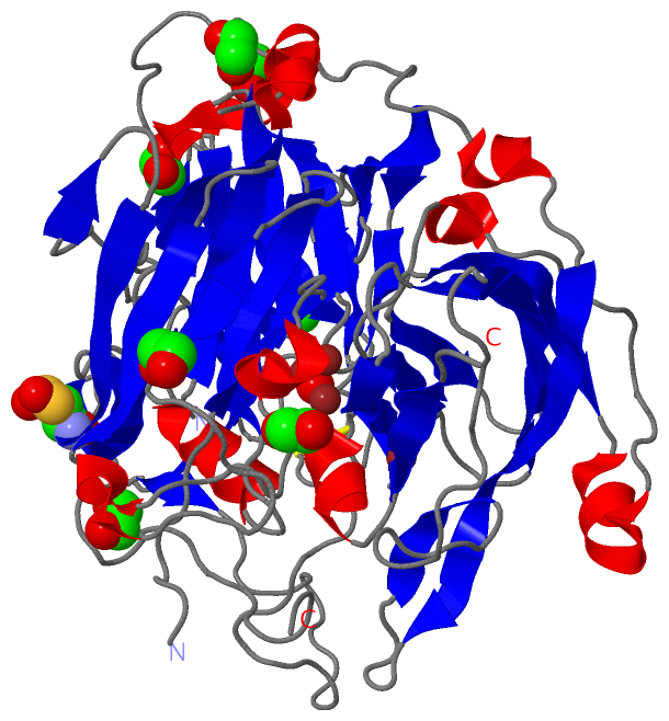
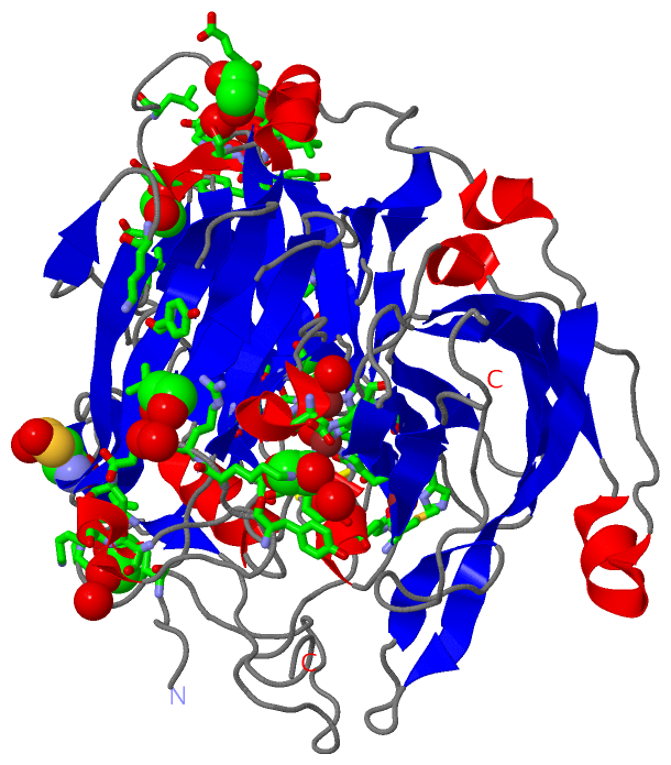
(-) Ligands, Modified Residues, Ions  (4, 13)

Asymmetric/Biological Unit (4, 13)
No.NameCountTypeFull Name
1CSX1Mod. Amino AcidS-OXY CYSTEINE
2CU4Ligand/IonCOPPER (II) ION
3EDO7Ligand/Ion1,2-ETHANEDIOL
4OH1Ligand/IonHYDROXIDE ION

(-) Sites  (12, 12)

Asymmetric Unit (12, 12)
No.NameEvidenceResiduesDescription
01AC1SOFTWAREHIS A:419 , CYS A:492 , HIS A:497 , MET A:502BINDING SITE FOR RESIDUE CU A 601
02AC2SOFTWAREHIS A:107 , HIS A:153 , HIS A:493 , OH A:605BINDING SITE FOR RESIDUE CU A 602
03AC3SOFTWAREHIS A:155 , HIS A:422 , HIS A:424 , HIS A:491 , CU A:604 , OH A:605BINDING SITE FOR RESIDUE CU A 603
04AC4SOFTWAREHIS A:105 , HIS A:107 , HIS A:422 , HIS A:424 , CU A:603 , OH A:605 , HOH A:2198BINDING SITE FOR RESIDUE CU A 604
05AC5SOFTWAREHIS A:105 , HIS A:107 , HIS A:153 , HIS A:155 , HIS A:422 , HIS A:424 , HIS A:491 , HIS A:493 , CU A:602 , CU A:603 , CU A:604BINDING SITE FOR RESIDUE OH A 605
06AC6SOFTWAREASP A:113 , TYR A:118 , TYR A:133 , LYS A:135 , HOH A:2575BINDING SITE FOR RESIDUE EDO A 701
07AC7SOFTWARELYS A:25 , TYR A:69 , THR A:307 , ALA A:308 , TYR A:309 , GLU A:310 , HOH A:2576BINDING SITE FOR RESIDUE EDO A 702
08AC8SOFTWAREASN A:74 , LEU A:76 , PRO A:77 , VAL A:100 , SER A:124 , LYS A:125 , HOH A:2577BINDING SITE FOR RESIDUE EDO A 703
09AC9SOFTWAREILE A:12 , GLU A:188 , GLU A:244 , TYR A:250BINDING SITE FOR RESIDUE EDO A 704
10BC1SOFTWAREARG A:136 , GLU A:137 , VAL A:138 , PRO A:247 , LEU A:343 , HOH A:2578 , HOH A:2579BINDING SITE FOR RESIDUE EDO A 705
11BC2SOFTWAREASP A:268 , PRO A:328 , ALA A:332 , ASN A:333 , HOH A:2580 , HOH A:2581BINDING SITE FOR RESIDUE EDO A 706
12BC3SOFTWARELEU A:184 , SER A:186 , GLU A:348 , HOH A:2582BINDING SITE FOR RESIDUE EDO A 707

(-) SS Bonds  (1, 1)

Asymmetric/Biological Unit
No.Residues
1A:229 -A:322

(-) Cis Peptide Bonds  (4, 4)

Asymmetric/Biological Unit
No.Residues
1Phe A:55 -Pro A:56
2Asn A:221 -Pro A:222
3Gly A:482 -Pro A:483
4Tyr A:500 -Asp A:501

 Sequence-Structure Mapping

(-) SAPs(SNPs)/Variants  (0, 0)

(no "SAP(SNP)/Variant" information available for 2X87)

(-) PROSITE Motifs  (0, 0)

(no "PROSITE Motif" information available for 2X87)

(-) Exons   (0, 0)

(no "Exon" information available for 2X87)

(-) Sequences/Alignments

Asymmetric/Biological Unit
   Reformat: Number of residues per line =  ('0' or empty: single-line sequence representation)
  Number of residues per labelling interval =   
  UniProt sequence: complete  aligned part    
   Show mapping: SCOP domains CATH domains Pfam domains Secondary structure (by author)
SAPs(SNPs) PROSITE motifs Exons
(details for a mapped element are shown in a popup box when the mouse pointer rests over it)
Chain A from PDB  Type:PROTEIN  Length:505
 aligned with COTA_BACSU | P07788 from UniProtKB/Swiss-Prot  Length:513

    Alignment length:510
                                    11        21        31        41        51        61        71        81        91       101       111       121       131       141       151       161       171       181       191       201       211       221       231       241       251       261       271       281       291       301       311       321       331       341       351       361       371       381       391       401       411       421       431       441       451       461       471       481       491       501       511
           COTA_BACSU     2 TLEKFVDALPIPDTLKPVQQSKEKTYYEVTMEECTHQLHRDLPPTRLWGYNGLFPGPTIEVKRNENVYVKWMNNLPSTHFLPIDHTIHHSDSQHEEPEVKTVVHLHGGVTPDDSDGYPEAWFSKDFEQTGPYFKREVYHYPNQQRGAILWYHDHAMALTRLNVYAGLVGAYIIHDPKEKRLKLPSDEYDVPLLITDRTINEDGSLFYPSAPENPSPSLPNPSIVPAFCGETILVNGKVWPYLEVEPRKYRFRVINASNTRTYNLSLDNGGDFIQIGSDGGLLPRSVKLNSFSLAPAERYDIIIDFTAYEGESIILANSAGCGGDVNPETDANIMQFRVTKPLAQKDESRKPKYLASYPSVQHERIQNIRTLKLAGTQDEYGRPVLLLNNKRWHDPVTETPKVGTTEIWSIINPTRGTHPIHLHLVSFRVLDRRPFDIARYQESGELSYTGPAVPPPPSEKGWKDTIQAHAGEVLRIAATFGPYSGRYVWHCHILEHEDYDMMRPMDITDP 511
               SCOP domains d2x87a1 A:2-182 Spore coat protein A, CotA                                                                                                                                           d2x87a2 A:183-356 Spore coat protein A, CotA                                                                                                                                  d2x87a3 A:357-511 Spore coat protein A, CotA                                                                                                                SCOP domains
               CATH domains ------------------------------------------------------------------------------------------------------------------------------------------------------------------------------------------------------------------------------------------------------------------------------------------------------------------------------------------------------------------------------------------------------------------------------------------------------------------------------------------------------------------------------ CATH domains
           Pfam domains (1) -----------------------------------------------------------------------------------------------------------------------------------------------------------------------------Cu-oxidase-2x87A01 A:175-340                                                                                                                                          -----------------Cu-oxidase_2-2x87A02 A:358-511                                                                                                                             Pfam domains (1)
           Pfam domains (2) ----------------------------------------------------------------------------------------------Cu-oxidase_3-2x87A03 A:96-179                                                       -------------------------------------------------------------------------------------------------------------------------------------------------------------------------------------------------------------------------------------------------------------------------------------------------------------------------------------------- Pfam domains (2)
           Pfam domains (3) ----------------------------------------------------------------------------------------------Cu-oxidase_3-2x87A04 A:96-179                                                       -------------------------------------------------------------------------------------------------------------------------------------------------------------------------------------------------------------------------------------------------------------------------------------------------------------------------------------------- Pfam domains (3)
         Sec.struct. author ..................ee....eeeeeeeeeeee........eeeeee........eeee....eeeeeee................-----........eeee....hhhhh.......hhhhh.........eeee......eeeeeee....hhhhhhhhh.eeeeeeehhhhhhhh..hhh.eeeeeeeeeee.....ee........................eeee..ee..eee....eeeeeeee......eeeee.....eeeeee..eeeeeeeee.eeee....eeeeeee.hhhh..eeeeee..............eeeeee...................hhhhh...eeeeeeeeeeeee.....eeeee................eeeeeeeee.....eeeee....eeeeeeee.hhhhhhhhh...........hhhhh....eeee...eeeeeeeee....eeeeeee.hhhhhh...eeeeee... Sec.struct. author
                 SAPs(SNPs) ------------------------------------------------------------------------------------------------------------------------------------------------------------------------------------------------------------------------------------------------------------------------------------------------------------------------------------------------------------------------------------------------------------------------------------------------------------------------------------------------------------------------------ SAPs(SNPs)
                    PROSITE ------------------------------------------------------------------------------------------------------------------------------------------------------------------------------------------------------------------------------------------------------------------------------------------------------------------------------------------------------------------------------------------------------------------------------------------------------------------------------------------------------------------------------ PROSITE
                 Transcript ------------------------------------------------------------------------------------------------------------------------------------------------------------------------------------------------------------------------------------------------------------------------------------------------------------------------------------------------------------------------------------------------------------------------------------------------------------------------------------------------------------------------------ Transcript
                 2x87 A   2 TLEKFVDALPIPDTLKPVQQSKEKTYYEVTMEEcTHQLHRDLPPTRLWGYNGLFPGPTIEVKRNENVYVKWMNNLPSTHFLPIDHTIHH-----EEPEVKTVVHLHGGVTPDDSDGYPEAWFSKDFEQTGPYFKREVYHYPNQQRGAILWYHDHAMALTRLNVYAGLVGAYIIHDPKEKRLKLPSDEYDVPLLITDRTINEDGSLFYPSAPENPSPSLPNPSIVPAFCGETILVNGKVWPYLEVEPRKYRFRVINASNTRTYNLSLDNGGDFIQIGSDGGLLPRSVKLNSFSLAPAERYDIIIDFTAYEGESIILANSAGCGGDVNPETDANIMQFRVTKPLAQKDESRKPKYLASYPSVQHERIQNIRTLKLAGTQDEYGRPVLLLNNKRWHDPVTETPKVGTTEIWSIINPTRGTHPIHLHLVSFRVLDRRPFDIARYQESGELSYTGPAVPPPPSEKGWKDTIQAHAGEVLRIAATFGPYSGRYVWHCHILEHEDYDMMRPMDITDP 511
                                    11        21        31   |    41        51        61        71        81        |-    |  101       111       121       131       141       151       161       171       181       191       201       211       221       231       241       251       261       271       281       291       301       311       321       331       341       351       361       371       381       391       401       411       421       431       441       451       461       471       481       491       501       511
                                                            35-CSX                                                 90    96                                                                                                                                                                                                                                                                                                                                                                                                                               

   Legend:   → Mismatch (orange background)
  - → Gap (green background, '-', border residues have a numbering label)
    → Modified Residue (blue background, lower-case, 'x' indicates undefined single-letter code, labelled with number + name)
  x → Chemical Group (purple background, 'x', labelled with number + name, e.g. ACE or NH2)
  extra numbering lines below/above indicate numbering irregularities and modified residue names etc., number ends below/above '|'

 Classification and Annotation

(-) SCOP Domains  (1, 3)

Asymmetric/Biological Unit

(-) CATH Domains  (0, 0)

(no "CATH Domain" information available for 2X87)

(-) Pfam Domains  (3, 4)

Asymmetric/Biological Unit

(-) Gene Ontology  (7, 7)

Asymmetric/Biological Unit(hide GO term definitions)
Chain A   (COTA_BACSU | P07788)
molecular function
    GO:0005507    copper ion binding    Interacting selectively and non-covalently with copper (Cu) ions.
    GO:0052716    hydroquinone:oxygen oxidoreductase activity    Catalysis of the reaction: 4 hydroquinone + O2 = 4 benzosemiquinone + 4 H2O.
    GO:0016491    oxidoreductase activity    Catalysis of an oxidation-reduction (redox) reaction, a reversible chemical reaction in which the oxidation state of an atom or atoms within a molecule is altered. One substrate acts as a hydrogen or electron donor and becomes oxidized, while the other acts as hydrogen or electron acceptor and becomes reduced.
    GO:0016722    oxidoreductase activity, oxidizing metal ions    Catalysis of an oxidation-reduction in which the oxidation state of metal ion is altered.
biological process
    GO:0055114    oxidation-reduction process    A metabolic process that results in the removal or addition of one or more electrons to or from a substance, with or without the concomitant removal or addition of a proton or protons.
    GO:0030435    sporulation resulting in formation of a cellular spore    The process in which a relatively unspecialized cell acquires the specialized features of a cellular spore, a cell form that can be used for dissemination, for survival of adverse conditions because of its heat and dessication resistance, and/or for reproduction.
cellular component
    GO:0030288    outer membrane-bounded periplasmic space    The region between the inner (cytoplasmic or plasma) membrane and outer membrane of organisms with two membranes such as Gram negative bacteria. These periplasmic spaces are relatively thick and contain a thin peptidoglycan layer (PGL), also referred to as a thin cell wall.

 Visualization

(-) Interactive Views

Asymmetric/Biological Unit
  Complete Structure
    Jena3D(integrated viewing of ligand, site, SAP, PROSITE, SCOP information)
    WebMol | AstexViewer[tm]@PDBe
(Java Applets, require no local installation except for Java; loading may be slow)
    STRAP
(Java WebStart application, automatic local installation, requires Java; full application with system access!)
    RasMol
(require local installation)
    Molscript (VRML)
(requires installation of a VRML viewer; select preferred view via VRML and generate a mono or stereo PDF format file)
 
  Ligands, Modified Residues, Ions
    CSX  [ RasMol | Jena3D ]  +environment [ RasMol | Jena3D ]
    CU  [ RasMol | Jena3D ]  +environment [ RasMol | Jena3D ]
    EDO  [ RasMol | Jena3D ]  +environment [ RasMol | Jena3D ]
    OH  [ RasMol | Jena3D ]  +environment [ RasMol | Jena3D ]
 
  Sites
    AC1  [ RasMol ]  +environment [ RasMol ]
    AC2  [ RasMol ]  +environment [ RasMol ]
    AC3  [ RasMol ]  +environment [ RasMol ]
    AC4  [ RasMol ]  +environment [ RasMol ]
    AC5  [ RasMol ]  +environment [ RasMol ]
    AC6  [ RasMol ]  +environment [ RasMol ]
    AC7  [ RasMol ]  +environment [ RasMol ]
    AC8  [ RasMol ]  +environment [ RasMol ]
    AC9  [ RasMol ]  +environment [ RasMol ]
    BC1  [ RasMol ]  +environment [ RasMol ]
    BC2  [ RasMol ]  +environment [ RasMol ]
    BC3  [ RasMol ]  +environment [ RasMol ]
 
  Cis Peptide Bonds
    Asn A:221 - Pro A:222   [ RasMol ]  
    Gly A:482 - Pro A:483   [ RasMol ]  
    Phe A:55 - Pro A:56   [ RasMol ]  
    Tyr A:500 - Asp A:501   [ RasMol ]  
 

(-) Still Images

Jmol
  protein: cartoon or spacefill or dots and stick; nucleic acid: cartoon and stick; ligands: spacefill; active site: stick
Molscript
  protein, nucleic acid: cartoon; ligands: spacefill; active site: ball and stick

 Databases and Analysis Tools

(-) Databases

Access by PDB/NDB ID
  2x87
    Family and Domain InformationProDom | SYSTERS
    General Structural InformationGlycoscienceDB | MMDB | NDB | OCA | PDB | PDBe | PDBj | PDBsum | PDBWiki | PQS | PROTEOPEDIA
    Orientation in MembranesOPM
    Protein SurfaceSURFACE
    Secondary StructureDSSP (structure derived) | HSSP (homology derived)
    Structural GenomicsGeneCensus
    Structural NeighboursCE | VAST
    Structure ClassificationCATH | Dali | SCOP
    Validation and Original DataBMRB Data View | BMRB Restraints Grid | EDS | PROCHECK | RECOORD | WHAT_CHECK
 
Access by UniProt ID/Accession number
  COTA_BACSU | P07788
    Comparative Protein Structure ModelsModBase
    Genomic InformationEnsembl
    Protein-protein InteractionDIP
    Sequence, Family and Domain InformationInterPro | Pfam | SMART | UniProtKB/SwissProt
 
Access by Enzyme Classificator   (EC Number)
  (no 'Enzyme Classificator' available)
    General Enzyme InformationBRENDA | EC-PDB | Enzyme | IntEnz
    PathwayKEGG | MetaCyc
 
Access by Disease Identifier   (MIM ID)
  (no 'MIM ID' available)
    Disease InformationOMIM
 
Access by GenAge ID
  (no 'GenAge ID' available)
    Age Related InformationGenAge

(-) Analysis Tools

Access by PDB/NDB ID
    Domain InformationXDom
    Interatomic Contacts of Structural UnitsCSU
    Ligand-protein ContactsLPC
    Protein CavitiescastP
    Sequence and Secondary StructurePDBCartoon
    Structure AlignmentSTRAP(Java WebStart application, automatic local installation, requires Java; full application with system access!)
    Structure and Sequence BrowserSTING
 
Access by UniProt ID/Accession number
  COTA_BACSU | P07788
    Protein Disorder PredictionDisEMBL | FoldIndex | GLOBPLOT (for more information see DisProt)

 Related Entries

(-) Entries Sharing at Least One Protein Chain (UniProt ID)

UniProtKB/Swiss-Prot
        COTA_BACSU | P077881gsk 1of0 1w6l 1w6w 1w8e 2bhf 2wsd 2x88 3zdw 4a66 4a67 4a68 4ako 4akp 4akq 4q89 4q8b 4yvn 4yvu

(-) Related Entries Specified in the PDB File

1gsk CRYSTAL STRUCTURE OF COTA, AN ENDOSPORE COAT PROTEIN FROM BACILLUS SUBTILIS
1of0 CRYSTAL STRUCTURE OF BACILLUS SUBTILIS COTA AFTER 1H SOAKING WITH ABTS
1uvw BACILLUS SUBTILIS COTA LACCASE ADDUCT WITH ABTS
1w6l 3D STRUCTURE OF COTA INCUBATED WITH CUCL2
1w6w 3D STRUCTURE OF COTA INCUBATED WITH SODIUM AZIDE
1w8e 3D STRUCTURE OF COTA INCUBATED WITH HYDROGEN PEROXIDE
2bhf 3D STRUCTURE OF THE REDUCED FORM OF COTA
2x88 CRYSTAL STRUCTURE OF HOLOCOTA