|
|
|
|
 Description
|
|
Compounds
|
||||||||||||||||||||||||||||||||||||||||
Chains, Units
Summary Information (see also Sequences/Alignments below) |
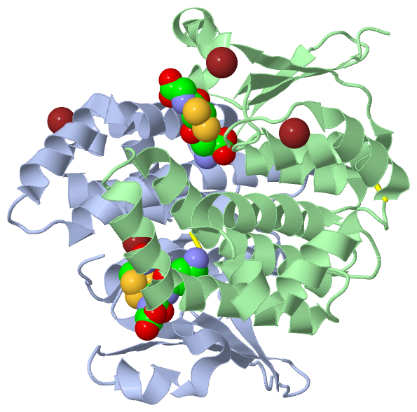
Ligands, Modified Residues, Ions (3, 8)
Asymmetric Unit (3, 8)
|
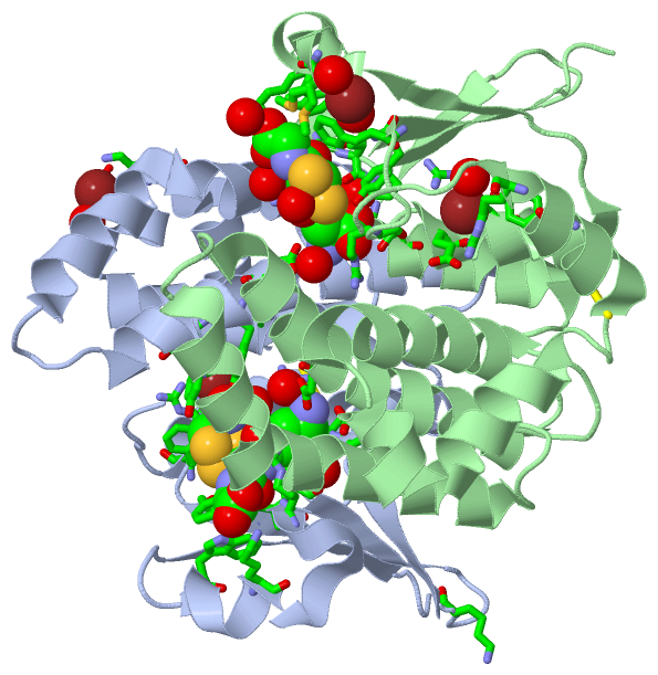
Sites (8, 8)
Asymmetric Unit (8, 8)
|
SS Bonds (2, 2)
Asymmetric Unit
|
||||||||||||
Cis Peptide Bonds (2, 2)
Asymmetric Unit
|
||||||||||||
SAPs(SNPs)/Variants (0, 0)| (no "SAP(SNP)/Variant" information available for 2WB9) |
PROSITE Motifs (0, 0)| (no "PROSITE Motif" information available for 2WB9) |
Exons (0, 0)| (no "Exon" information available for 2WB9) |
Sequences/Alignments
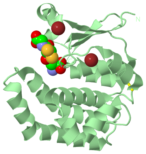
Asymmetric UnitChain A from PDB Type:PROTEIN Length:210 aligned with Q06A71_FASHE | Q06A71 from UniProtKB/TrEMBL Length:211 Alignment length:210 11 21 31 41 51 61 71 81 91 101 111 121 131 141 151 161 171 181 191 201 211 Q06A71_FASHE 2 DKQHFKLWYFQFRGRAEPIRLLLTCAGVKFEDYQFTMDQWPTIKPTLPGGRVPLLDVTGPDGKLRRYQESMAIARLLARQFKMMGETDEEYYLIERIIGECEDLYREVYTIFRTPQGEKEAKIKEFKENNGPTLLKLVSESLESSGGKHVAGNRITLGDLFLFTTLTHVMETVPGFLEQKFPKLHEFHKSLPTSCSRLSEYLKKRAKTPF 211 SCOP domains d2wb9a1 A:2-84 automated matches d2wb9a2 A:85-211 automated matches SCOP domains CATH domains 2wb9A01 A:2-83 Glutaredoxin 2wb9A02 A:84-211 [code=1.20.1050.10, no name defined] CATH domains Pfam domains ------------------------------------------------------------------------------------------------------------------------------------------------------------------------------------------------------------------ Pfam domains SAPs(SNPs) ------------------------------------------------------------------------------------------------------------------------------------------------------------------------------------------------------------------ SAPs(SNPs) PROSITE ------------------------------------------------------------------------------------------------------------------------------------------------------------------------------------------------------------------ PROSITE Transcript ------------------------------------------------------------------------------------------------------------------------------------------------------------------------------------------------------------------ Transcript 2wb9 A 2 DKQHFKLWYFQFRGRAEPIRLLLTCAGVKFEDYQFTMDQWPTIKPTLPGGRVPLLDVTGPDGKLRRYQESMAIARLLARQFKMMGETDEEYYLIERIIGECEDLYREVYTIFRTPQGEKEAKIKEFKENNGPTLLKLVSESLESSGGKHVAGNRITLGDLFLFTTLTHVMETVPGFLEQKFPKLHEFHKSLPTSCSRLSEYLKKRAKTPF 211 11 21 31 41 51 61 71 81 91 101 111 121 131 141 151 161 171 181 191 201 211 Chain B from PDB Type:PROTEIN Length:210 aligned with Q06A71_FASHE | Q06A71 from UniProtKB/TrEMBL Length:211 Alignment length:210 11 21 31 41 51 61 71 81 91 101 111 121 131 141 151 161 171 181 191 201 211 Q06A71_FASHE 2 DKQHFKLWYFQFRGRAEPIRLLLTCAGVKFEDYQFTMDQWPTIKPTLPGGRVPLLDVTGPDGKLRRYQESMAIARLLARQFKMMGETDEEYYLIERIIGECEDLYREVYTIFRTPQGEKEAKIKEFKENNGPTLLKLVSESLESSGGKHVAGNRITLGDLFLFTTLTHVMETVPGFLEQKFPKLHEFHKSLPTSCSRLSEYLKKRAKTPF 211 SCOP domains d2wb9b1 B:2-84 automated matches d2wb9b2 B:85-211 automated matches SCOP domains CATH domains 2wb9B01 B:2-83 Glutaredoxin 2wb9B02 B:84-211 [code=1.20.1050.10, no name defined] CATH domains Pfam domains (1) -----GST_N-2wb9B03 B:7-80 -----GST_C-2wb9B01 B:86-193 ------------------ Pfam domains (1) Pfam domains (2) -----GST_N-2wb9B04 B:7-80 -----GST_C-2wb9B02 B:86-193 ------------------ Pfam domains (2) SAPs(SNPs) ------------------------------------------------------------------------------------------------------------------------------------------------------------------------------------------------------------------ SAPs(SNPs) PROSITE ------------------------------------------------------------------------------------------------------------------------------------------------------------------------------------------------------------------ PROSITE Transcript ------------------------------------------------------------------------------------------------------------------------------------------------------------------------------------------------------------------ Transcript 2wb9 B 2 DKQHFKLWYFQFRGRAEPIRLLLTCAGVKFEDYQFTMDQWPTIKPTLPGGRVPLLDVTGPDGKLRRYQESMAIARLLARQFKMMGETDEEYYLIERIIGECEDLYREVYTIFRTPQGEKEAKIKEFKENNGPTLLKLVSESLESSGGKHVAGNRITLGDLFLFTTLTHVMETVPGFLEQKFPKLHEFHKSLPTSCSRLSEYLKKRAKTPF 211 11 21 31 41 51 61 71 81 91 101 111 121 131 141 151 161 171 181 191 201 211
|
||||||||||||||||||||
SCOP Domains (2, 4)
Asymmetric Unit
 |
CATH Domains (2, 4)
Asymmetric Unit
|
Pfam Domains (2, 4)
Asymmetric Unit
|
Gene Ontology (2, 2)|
Asymmetric Unit(hide GO term definitions) Chain A,B (Q06A71_FASHE | Q06A71)
|
||||||||||||||||||
Interactive Views
|
||||||||||||||||||||||||||||||||||||||||||||||||||||||||||||||||||||||||||||||||||||||||||||||||||||||||||||||||||||||||||||||||||||||||||||||||||||||||||||||||||||||||||||||||||||||||||||||||||||||||||||||||||||
Still Images
|
||||||||||||||||
Databases
|
||||||||||||||||||||||||||||||||||||||||||||||||||||||||||||||||||||||||||||||||||||||||||||||||||||||||||||||||||||||||||||||||||||||||||||||||||||||||||||||||
Analysis Tools
|
|||||||||||||||||||||||||||||||||||||||||||||||||||||||||||||
Entries Sharing at Least One Protein Chain (UniProt ID)
Related Entries Specified in the PDB File
|
|