| molecular function |
|---|
| | GO:0000175 | | 3'-5'-exoribonuclease activity | | Catalysis of the sequential cleavage of mononucleotides from a free 3' terminus of an RNA molecule. |
| | GO:0051575 | | 5'-deoxyribose-5-phosphate lyase activity | | Catalysis of the beta-elimination of the 5' deoxyribose-5-phosphate at an abasic site in DNA where a DNA-(apurinic or apyrimidinic site) lyase has already cleaved the C-O-P bond 3' to the apurinic or apyrimidinic site. |
| | GO:0003677 | | DNA binding | | Any molecular function by which a gene product interacts selectively and non-covalently with DNA (deoxyribonucleic acid). |
| | GO:0008852 | | exodeoxyribonuclease I activity | | Catalysis of the degradation of single-stranded DNA. It acts progressively in a 3' to 5' direction, releasing 5'-phosphomononucleotides. |
| | GO:0004529 | | exodeoxyribonuclease activity | | Catalysis of the sequential cleavage of mononucleotides from a free 5' or 3' terminus of a DNA molecule. |
| | GO:0004527 | | exonuclease activity | | Catalysis of the hydrolysis of ester linkages within nucleic acids by removing nucleotide residues from the 3' or 5' end. |
| | GO:0016787 | | hydrolase activity | | Catalysis of the hydrolysis of various bonds, e.g. C-O, C-N, C-C, phosphoric anhydride bonds, etc. Hydrolase is the systematic name for any enzyme of EC class 3. |
| | GO:0000287 | | magnesium ion binding | | Interacting selectively and non-covalently with magnesium (Mg) ions. |
| | GO:0046872 | | metal ion binding | | Interacting selectively and non-covalently with any metal ion. |
| | GO:0004518 | | nuclease activity | | Catalysis of the hydrolysis of ester linkages within nucleic acids. |
| | GO:0003676 | | nucleic acid binding | | Interacting selectively and non-covalently with any nucleic acid. |
| | GO:0005515 | | protein binding | | Interacting selectively and non-covalently with any protein or protein complex (a complex of two or more proteins that may include other nonprotein molecules). |
| | GO:0008310 | | single-stranded DNA 3'-5' exodeoxyribonuclease activity | | Catalysis of the sequential cleavage of mononucleotides from a free 3' terminus of a single-stranded DNA molecule. |
| | GO:0003697 | | single-stranded DNA binding | | Interacting selectively and non-covalently with single-stranded DNA. |
| biological process |
|---|
| | GO:0006308 | | DNA catabolic process | | The cellular DNA metabolic process resulting in the breakdown of DNA, deoxyribonucleic acid, one of the two main types of nucleic acid, consisting of a long unbranched macromolecule formed from one or two strands of linked deoxyribonucleotides, the 3'-phosphate group of each constituent deoxyribonucleotide being joined in 3',5'-phosphodiester linkage to the 5'-hydroxyl group of the deoxyribose moiety of the next one. |
| | GO:0000738 | | DNA catabolic process, exonucleolytic | | The chemical reactions and pathways resulting in the breakdown of DNA, involving the hydrolysis of terminal 3',5'-phosphodiester bonds in one or two strands of deoxyribonucleotides. |
| | GO:0006281 | | DNA repair | | The process of restoring DNA after damage. Genomes are subject to damage by chemical and physical agents in the environment (e.g. UV and ionizing radiations, chemical mutagens, fungal and bacterial toxins, etc.) and by free radicals or alkylating agents endogenously generated in metabolism. DNA is also damaged because of errors during its replication. A variety of different DNA repair pathways have been reported that include direct reversal, base excision repair, nucleotide excision repair, photoreactivation, bypass, double-strand break repair pathway, and mismatch repair pathway. |
| | GO:0090503 | | RNA phosphodiester bond hydrolysis, exonucleolytic | | The chemical reactions and pathways involving the hydrolysis of terminal 3',5'-phosphodiester bonds in one or two strands of ribonucleotides. |
| | GO:0006974 | | cellular response to DNA damage stimulus | | Any process that results in a change in state or activity of a cell (in terms of movement, secretion, enzyme production, gene expression, etc.) as a result of a stimulus indicating damage to its DNA from environmental insults or errors during metabolism. |
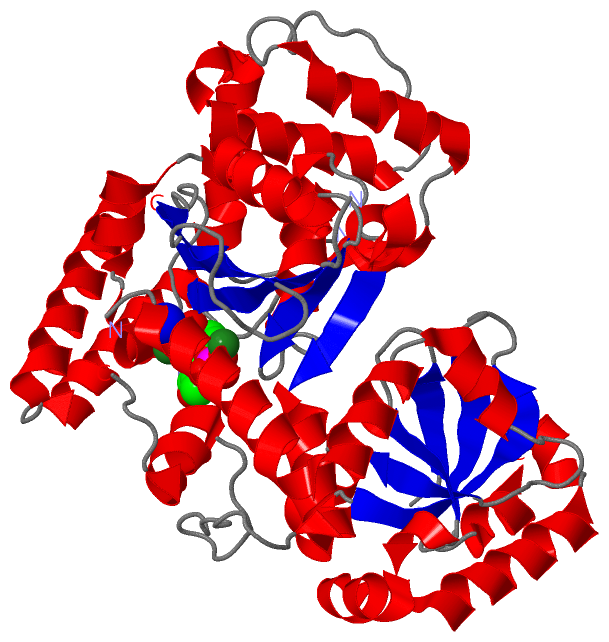
 Description
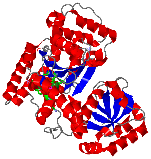