|
|
|
|
 Description
|
|
Compounds
|
||||||||||||||||||||||||||||||||||||||||
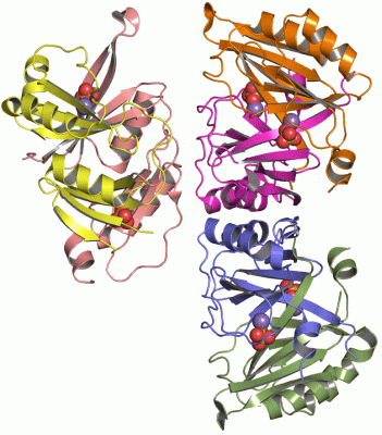
Chains, Units
Summary Information (see also Sequences/Alignments below) |
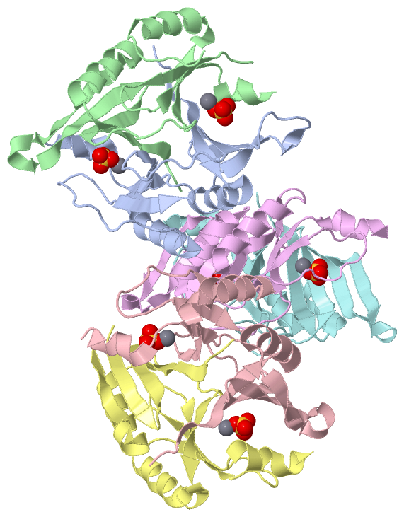
Ligands, Modified Residues, Ions (2, 12)| Asymmetric Unit (2, 12) Biological Unit 1 (1, 2) Biological Unit 2 (1, 2) Biological Unit 3 (1, 2) Biological Unit 4 (1, 2) |
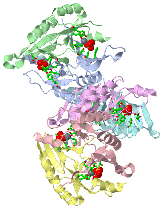
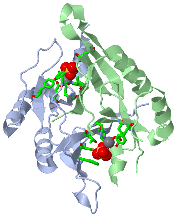
Sites (12, 12)
Asymmetric Unit (12, 12)
|
SS Bonds (0, 0)| (no "SS Bond" information available for 2P7P) |
Cis Peptide Bonds (0, 0)| (no "Cis Peptide Bond" information available for 2P7P) |
SAPs(SNPs)/Variants (0, 0)| (no "SAP(SNP)/Variant" information available for 2P7P) |
PROSITE Motifs (1, 6)
Asymmetric Unit (1, 6)
|
||||||||||||||||||||||||||||||||||||||||||||||||||||||||||||||||||||||||||||||||||||||||||||||||||||||||||||||||||||||||
Exons (0, 0)| (no "Exon" information available for 2P7P) |
Sequences/Alignments
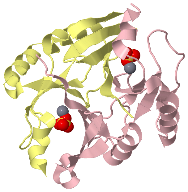
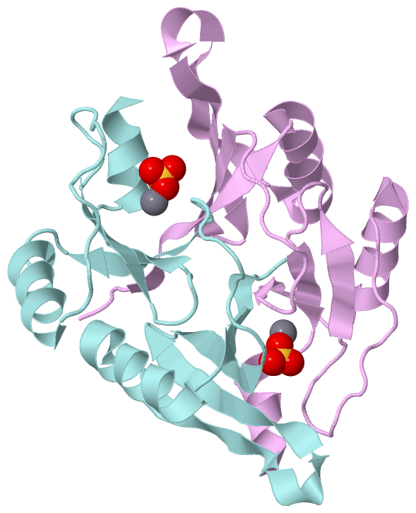
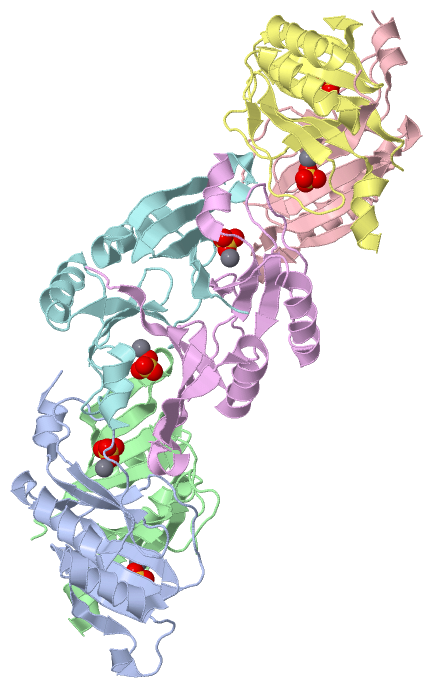
Asymmetric UnitChain A from PDB Type:PROTEIN Length:131 aligned with FOSX_LISMO | Q8Y6I2 from UniProtKB/Swiss-Prot Length:133 Alignment length:131 10 20 30 40 50 60 70 80 90 100 110 120 130 FOSX_LISMO 1 MISGLSHITLIVKDLNKTTTFLREIFNAEEIYSSGDQTFSLSKEKFFLIAGLWICIMEGDSLQEQTYNHIAFRIQSEEVDEYIERIKSLGVEIKPERPRVEGEGRSIYFYDFDNHLFELHAGTLEERLKRY 131 SCOP domains d2p7pa_ A: automated matches SCOP domains CATH domains 2p7pA00 A:1-131 2,3-Dihydroxybiphenyl 1,2-Dioxygenase, domain 1 CATH domains Pfam domains ----------------------------------------------------------------------------------------------------------------------------------- Pfam domains SAPs(SNPs) ----------------------------------------------------------------------------------------------------------------------------------- SAPs(SNPs) PROSITE ---VOC PDB: A:4-122 UniProt: 4-122 --------- PROSITE Transcript ----------------------------------------------------------------------------------------------------------------------------------- Transcript 2p7p A 1 MISGLSHITLIVKDLNKTTAFLQNIFNAEEIYSSGDKTFSLSKEKFFLIAGLWICIMEGDSLQERTYNHIAFQIQSEEVDEYTERIKALGVEMKPERPRVQGEGRSIYFYDFDNHLFELHAGTLEERLKRY 131 10 20 30 40 50 60 70 80 90 100 110 120 130 Chain B from PDB Type:PROTEIN Length:122 aligned with FOSX_LISMO | Q8Y6I2 from UniProtKB/Swiss-Prot Length:133 Alignment length:129 10 20 30 40 50 60 70 80 90 100 110 120 FOSX_LISMO 1 MISGLSHITLIVKDLNKTTTFLREIFNAEEIYSSGDQTFSLSKEKFFLIAGLWICIMEGDSLQEQTYNHIAFRIQSEEVDEYIERIKSLGVEIKPERPRVEGEGRSIYFYDFDNHLFELHAGTLEERLK 129 SCOP domains d2p7pb_ B: automated matches SCOP domains CATH domains 2p7pB00 B:1-129 2,3-Dihydroxybiphenyl 1,2-Dioxygenase, domain 1 CATH domains Pfam domains --------------------------------------------------------------------------------------------------------------------------------- Pfam domains SAPs(SNPs) --------------------------------------------------------------------------------------------------------------------------------- SAPs(SNPs) PROSITE ---VOC PDB: B:4-122 UniProt: 4-122 ------- PROSITE Transcript --------------------------------------------------------------------------------------------------------------------------------- Transcript 2p7p B 1 MISGLSHITLIVKDLNKTTAFLQNIFNAEEIYSSGDKTFSLSKEKFFLIAGLWICIMEGDSLQERTYNHIAFQIQSEEVDEYTERIKALGVEMKP-------EGRSIYFYDFDNHLFELHAGTLEERLK 129 10 20 30 40 50 60 70 80 90 | - | 110 120 95 103 Chain C from PDB Type:PROTEIN Length:131 aligned with FOSX_LISMO | Q8Y6I2 from UniProtKB/Swiss-Prot Length:133 Alignment length:131 10 20 30 40 50 60 70 80 90 100 110 120 130 FOSX_LISMO 1 MISGLSHITLIVKDLNKTTTFLREIFNAEEIYSSGDQTFSLSKEKFFLIAGLWICIMEGDSLQEQTYNHIAFRIQSEEVDEYIERIKSLGVEIKPERPRVEGEGRSIYFYDFDNHLFELHAGTLEERLKRY 131 SCOP domains d2p7pc_ C: automated matches SCOP domains CATH domains 2p7pC00 C:1-131 2,3-Dihydroxybiphenyl 1,2-Dioxygenase, domain 1 CATH domains Pfam domains ----------------------------------------------------------------------------------------------------------------------------------- Pfam domains SAPs(SNPs) ----------------------------------------------------------------------------------------------------------------------------------- SAPs(SNPs) PROSITE ---VOC PDB: C:4-122 UniProt: 4-122 --------- PROSITE Transcript ----------------------------------------------------------------------------------------------------------------------------------- Transcript 2p7p C 1 MISGLSHITLIVKDLNKTTAFLQNIFNAEEIYSSGDKTFSLSKEKFFLIAGLWICIMEGDSLQERTYNHIAFQIQSEEVDEYTERIKALGVEMKPERPRVQGEGRSIYFYDFDNHLFELHAGTLEERLKRY 131 10 20 30 40 50 60 70 80 90 100 110 120 130 Chain D from PDB Type:PROTEIN Length:125 aligned with FOSX_LISMO | Q8Y6I2 from UniProtKB/Swiss-Prot Length:133 Alignment length:132 10 20 30 40 50 60 70 80 90 100 110 120 130 FOSX_LISMO 1 MISGLSHITLIVKDLNKTTTFLREIFNAEEIYSSGDQTFSLSKEKFFLIAGLWICIMEGDSLQEQTYNHIAFRIQSEEVDEYIERIKSLGVEIKPERPRVEGEGRSIYFYDFDNHLFELHAGTLEERLKRYH 132 SCOP domains d2p7pd_ D: automated matches SCOP domains CATH domains 2p7pD00 D:1-132 2,3-Dihydroxybiphe nyl 1,2-Dioxygenase, domain 1 CATH domains Pfam domains ------------------------------------------------------------------------------------------------------------------------------------ Pfam domains SAPs(SNPs) ------------------------------------------------------------------------------------------------------------------------------------ SAPs(SNPs) PROSITE ---VOC PDB: D:4-122 UniProt: 4-122 ---------- PROSITE Transcript ------------------------------------------------------------------------------------------------------------------------------------ Transcript 2p7p D 1 MISGLSHITLIVKDLNKTTAFLQNIFNAEEIYSS-------SKEKFFLIAGLWICIMEGDSLQERTYNHIAFQIQSEEVDEYTERIKALGVEMKPERPRVQGEGRSIYFYDFDNHLFELHAGTLEERLKRYH 132 10 20 30 | - | 50 60 70 80 90 100 110 120 130 34 42 Chain E from PDB Type:PROTEIN Length:131 aligned with FOSX_LISMO | Q8Y6I2 from UniProtKB/Swiss-Prot Length:133 Alignment length:131 10 20 30 40 50 60 70 80 90 100 110 120 130 FOSX_LISMO 1 MISGLSHITLIVKDLNKTTTFLREIFNAEEIYSSGDQTFSLSKEKFFLIAGLWICIMEGDSLQEQTYNHIAFRIQSEEVDEYIERIKSLGVEIKPERPRVEGEGRSIYFYDFDNHLFELHAGTLEERLKRY 131 SCOP domains d2p7pe_ E: automated matches SCOP domains CATH domains 2p7pE00 E:1-131 2,3-Dihydroxybiphenyl 1,2-Dioxygenase, domain 1 CATH domains Pfam domains ----------------------------------------------------------------------------------------------------------------------------------- Pfam domains SAPs(SNPs) ----------------------------------------------------------------------------------------------------------------------------------- SAPs(SNPs) PROSITE ---VOC PDB: E:4-122 UniProt: 4-122 --------- PROSITE Transcript ----------------------------------------------------------------------------------------------------------------------------------- Transcript 2p7p E 1 MISGLSHITLIVKDLNKTTAFLQNIFNAEEIYSSGDKTFSLSKEKFFLIAGLWICIMEGDSLQERTYNHIAFQIQSEEVDEYTERIKALGVEMKPERPRVQGEGRSIYFYDFDNHLFELHAGTLEERLKRY 131 10 20 30 40 50 60 70 80 90 100 110 120 130 Chain F from PDB Type:PROTEIN Length:131 aligned with FOSX_LISMO | Q8Y6I2 from UniProtKB/Swiss-Prot Length:133 Alignment length:131 10 20 30 40 50 60 70 80 90 100 110 120 130 FOSX_LISMO 1 MISGLSHITLIVKDLNKTTTFLREIFNAEEIYSSGDQTFSLSKEKFFLIAGLWICIMEGDSLQEQTYNHIAFRIQSEEVDEYIERIKSLGVEIKPERPRVEGEGRSIYFYDFDNHLFELHAGTLEERLKRY 131 SCOP domains d2p7pf_ F: automated matches SCOP domains CATH domains 2p7pF00 F:1-131 2,3-Dihydroxybiphenyl 1,2-Dioxygenase, domain 1 CATH domains Pfam domains ----------------------------------------------------------------------------------------------------------------------------------- Pfam domains SAPs(SNPs) ----------------------------------------------------------------------------------------------------------------------------------- SAPs(SNPs) PROSITE ---VOC PDB: F:4-122 UniProt: 4-122 --------- PROSITE Transcript ----------------------------------------------------------------------------------------------------------------------------------- Transcript 2p7p F 1 MISGLSHITLIVKDLNKTTAFLQNIFNAEEIYSSGDKTFSLSKEKFFLIAGLWICIMEGDSLQERTYNHIAFQIQSEEVDEYTERIKALGVEMKPERPRVQGEGRSIYFYDFDNHLFELHAGTLEERLKRY 131 10 20 30 40 50 60 70 80 90 100 110 120 130
|
||||||||||||||||||||
SCOP Domains (1, 6)
Asymmetric Unit
 |
CATH Domains (1, 6)
Asymmetric Unit
|
Pfam Domains (0, 0)| (no "Pfam Domain" information available for 2P7P) |
Gene Ontology (3, 3)|
Asymmetric Unit(hide GO term definitions) Chain A,B,C,D,E,F (FOSX_LISMO | Q8Y6I2)
|
||||||||||||||||||||||||||||||||||||
Interactive Views
|
|||||||||||||||||||||||||||||||||||||||||||||||||||||||||||||||||||||||||||||||||||||||||||||||||||||||||||||||||||||||||||||||||||||||||||||||||||||||||||||||||||||||||||||||||||||||||||||||||||||||||||||||||||||||||||||||||||||||||||
Still Images
|
||||||||||||||||
Databases
|
||||||||||||||||||||||||||||||||||||||||||||||||||||||||||||||||||||||||||||||||||||||||||||||||||||||||||||||||||||||||||||||||||||||||||||||||||||||||||||||||
Analysis Tools
|
|||||||||||||||||||||||||||||||||||||||||||||||||||||||||||||
Entries Sharing at Least One Protein Chain (UniProt ID)
Related Entries Specified in the PDB File
|
|