| molecular function |
|---|
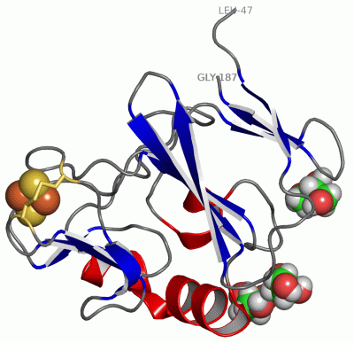
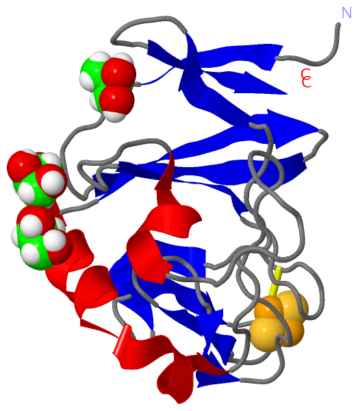
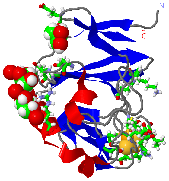
| | GO:0051537 | | 2 iron, 2 sulfur cluster binding | | Interacting selectively and non-covalently with a 2 iron, 2 sulfur (2Fe-2S) cluster; this cluster consists of two iron atoms, with two inorganic sulfur atoms found between the irons and acting as bridging ligands. |
| | GO:0051536 | | iron-sulfur cluster binding | | Interacting selectively and non-covalently with an iron-sulfur cluster, a combination of iron and sulfur atoms. |
| | GO:0046872 | | metal ion binding | | Interacting selectively and non-covalently with any metal ion. |
| | GO:0016491 | | oxidoreductase activity | | Catalysis of an oxidation-reduction (redox) reaction, a reversible chemical reaction in which the oxidation state of an atom or atoms within a molecule is altered. One substrate acts as a hydrogen or electron donor and becomes oxidized, while the other acts as hydrogen or electron acceptor and becomes reduced. |
| | GO:0016679 | | oxidoreductase activity, acting on diphenols and related substances as donors | | Catalysis of an oxidation-reduction (redox) reaction in which a diphenol or related substance acts as a hydrogen or electron donor and reduces a hydrogen or electron acceptor. |
| | GO:0008121 | | ubiquinol-cytochrome-c reductase activity | | Catalysis of the transfer of a solute or solutes from one side of a membrane to the other according to the reaction: CoQH2 + 2 ferricytochrome c = CoQ + 2 ferrocytochrome c + 2 H+. |
| biological process |
|---|
| | GO:1902600 | | hydrogen ion transmembrane transport | | The directed movement of hydrogen ion (proton) across a membrane. |
| | GO:0055114 | | oxidation-reduction process | | A metabolic process that results in the removal or addition of one or more electrons to or from a substance, with or without the concomitant removal or addition of a proton or protons. |
| cellular component |
|---|
| | GO:0016021 | | integral component of membrane | | The component of a membrane consisting of the gene products and protein complexes having at least some part of their peptide sequence embedded in the hydrophobic region of the membrane. |
| | GO:0016020 | | membrane | | A lipid bilayer along with all the proteins and protein complexes embedded in it an attached to it. |
| | GO:0005886 | | plasma membrane | | The membrane surrounding a cell that separates the cell from its external environment. It consists of a phospholipid bilayer and associated proteins. |
 Description