|
|
|
|
 Description
|
|
Compounds
|
||||||||||||||||||||||||||||||||||||||||||||||||||||||||||||||||||||||||||
Chains, Units
Summary Information (see also Sequences/Alignments below) |
Ligands, Modified Residues, Ions (2, 2)
Sites (2, 2)
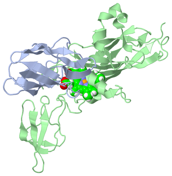
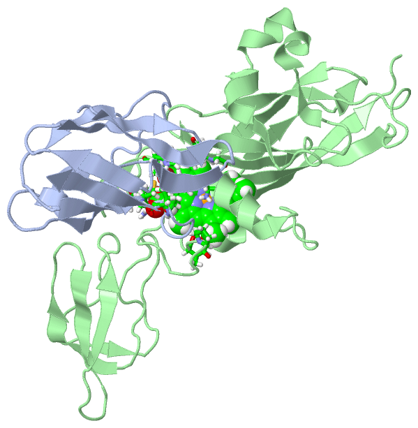
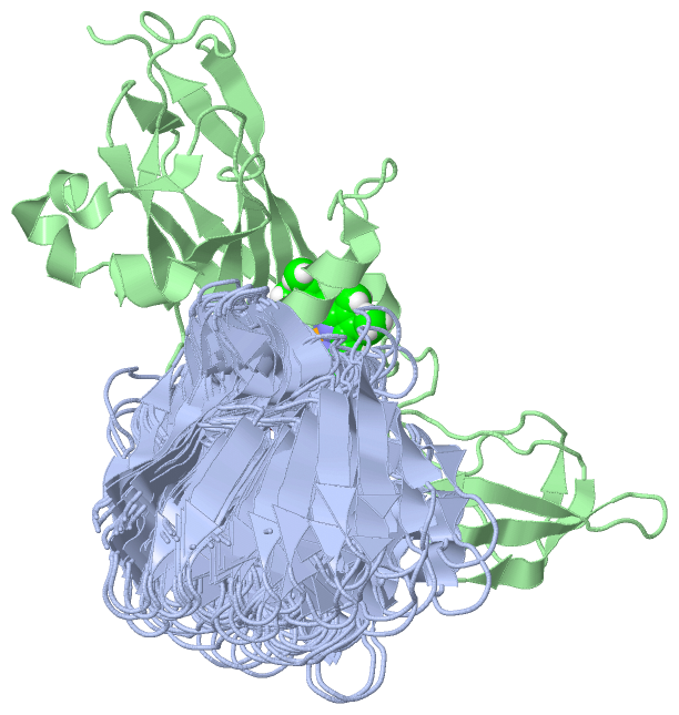
NMR Structure (2, 2)
|
SS Bonds (0, 0)| (no "SS Bond" information available for 2JXM) |
Cis Peptide Bonds (3, 60)
NMR Structure
|
||||||||||||||||||||
SAPs(SNPs)/Variants (0, 0)| (no "SAP(SNP)/Variant" information available for 2JXM) |
PROSITE Motifs (1, 1)
NMR Structure (1, 1)
|
||||||||||||||||||||||||
Exons (0, 0)| (no "Exon" information available for 2JXM) |
Sequences/Alignments
NMR StructureChain A from PDB Type:PROTEIN Length:97 aligned with PLAS_PROHO | P50057 from UniProtKB/Swiss-Prot Length:131 Alignment length:97 44 54 64 74 84 94 104 114 124 PLAS_PROHO 35 ATVQIKMGTDKYAPLYEPKALSISAGDTVEFVMNKVGPHNVIFDKVPAGESAPALSNTKLAIAPGSFYSVTLGTPGTYSFYCTPHRGAGMVGTITVE 131 SCOP domains d2jxma_ A: Plastocyanin SCOP domains CATH domains 2jxmA00 A:1-97 Cupredoxins - blue copper proteins CATH domains Pfam domains --Copper-bind-2jxmA01 A:3-97 Pfam domains SAPs(SNPs) ------------------------------------------------------------------------------------------------- SAPs(SNPs) PROSITE ---------------------------------------------------------------------------COPPER_BLUE ------- PROSITE Transcript ------------------------------------------------------------------------------------------------- Transcript 2jxm A 1 ASVQIKMGTDKYAPLYEPKALSISAGDTVEFVMNKVGPHNVIFDKVPAGESAPALSNTKLAIAPGSFYSVTLGTPGTYSFYCTPHRGAGMVGTITVE 97 10 20 30 40 50 60 70 80 90 Chain B from PDB Type:PROTEIN Length:249 aligned with Q8RN59_PROHO | Q8RN59 from UniProtKB/TrEMBL Length:562 Alignment length:249 56 66 76 86 96 106 116 126 136 146 156 166 176 186 196 206 216 226 236 246 256 266 276 286 Q8RN59_PROHO 47 YPFYAQYNYDSPREATGKIVCANCHLAKKTVEIEVPQAVLPDTVFKAVVKVPYDLDIQQVQADGSPSGLNVGAVLMLPEGFKLAPPERVDEELMEEVGDFYYLVTPYSETDENILLAGPLPGEDYQEMIFPILSPNPATDAGVYFGKYSIHLGGNRGRGQVYPTGELSNNNAFSASIAGTIAAIEDNGFGFDVTIQPEDGDAVVTSILPGPELIVAVGDTVEAGQLLTTNPNVGGFGQMDSEIVLQSSS 295 SCOP domains d2jxmb1 B:1-246 Cytochrome f-plastocyanin complex --- SCOP domains CATH domains 2jxmB01 B:1-172,B:230-248 [code=2.60.40.830, no name defined] 2jxmB02 B:173-229 [code=2.40.50.100, no name defined] 2jxmB01 - CATH domains Pfam domains ------------------------------------------------------------------------------------------------------------------------------------------------------------------------Apocytochr_F_C-2jxmB01 B:169-249 Pfam domains SAPs(SNPs) --------------------------------------------------------------------------------------------------------------------------------------------------------------------------------------------------------------------------------------------------------- SAPs(SNPs) PROSITE --------------------------------------------------------------------------------------------------------------------------------------------------------------------------------------------------------------------------------------------------------- PROSITE Transcript --------------------------------------------------------------------------------------------------------------------------------------------------------------------------------------------------------------------------------------------------------- Transcript 2jxm B 1 YPFYAQYNYDSPREATGKIVCANCHLAKKTVEIEVPQAVLPDTVFKAVVKVPYDLDIQQVQADGSPSGLNVGAVLMLPEGFKLAPPERVDEELMEEVGDFYYLVTPYSETDENILLAGPLPGEDYQEMIFPILSPNPATDAGVYFGKYSIHLGGNRGRGQVYPTGELSNNNAFSASIAGTIAAIEDNGFGFDVTIQPEDGDAVVTSILPGPELIVAVGDTVEAGQLLTTNPNVGGFGQMDSEIVLQSSS 249 10 20 30 40 50 60 70 80 90 100 110 120 130 140 150 160 170 180 190 200 210 220 230 240
|
||||||||||||||||||||
SCOP Domains (2, 2)
NMR Structure
 |
CATH Domains (3, 3)
NMR Structure
|
Pfam Domains (2, 2)
NMR Structure
|
Gene Ontology (12, 15)|
NMR Structure(hide GO term definitions) Chain A (PLAS_PROHO | P50057)
Chain B (Q8RN59_PROHO | Q8RN59)
|
||||||||||||||||||||||||||||||||||||||||||||||||||||||||||||||||||||||||||||||||||||||||||||||||||||||||||||||||||||||||||||||
Interactive Views
|
|||||||||||||||||||||||||||||||||||||||||||||||||||||||||||||||||||||||||||||||||||||||||||||||||||||||||||||||||||||||||||||||||||||||||||||||||||
Still Images
|
||||||||||||||||
Databases
|
||||||||||||||||||||||||||||||||||||||||||||||||||||||||||||||||||||||||||||||||||||||||||||||||||||||||||||||||||||||||||||||||||||||||||||||||||||||||||||||||||||||||||||||||||||||||||
Analysis Tools
|
||||||||||||||||||||||||||||||||||||||||||||||||||||||||||||||||||||||||
Entries Sharing at Least One Protein Chain (UniProt ID)
Related Entries Specified in the PDB File
|
|