|
|
|
|
 Description
|
|
Compounds
|
||||||||||||||||||||||||||||||||||||||||||||||||||||||||
Chains, Units
Summary Information (see also Sequences/Alignments below) |
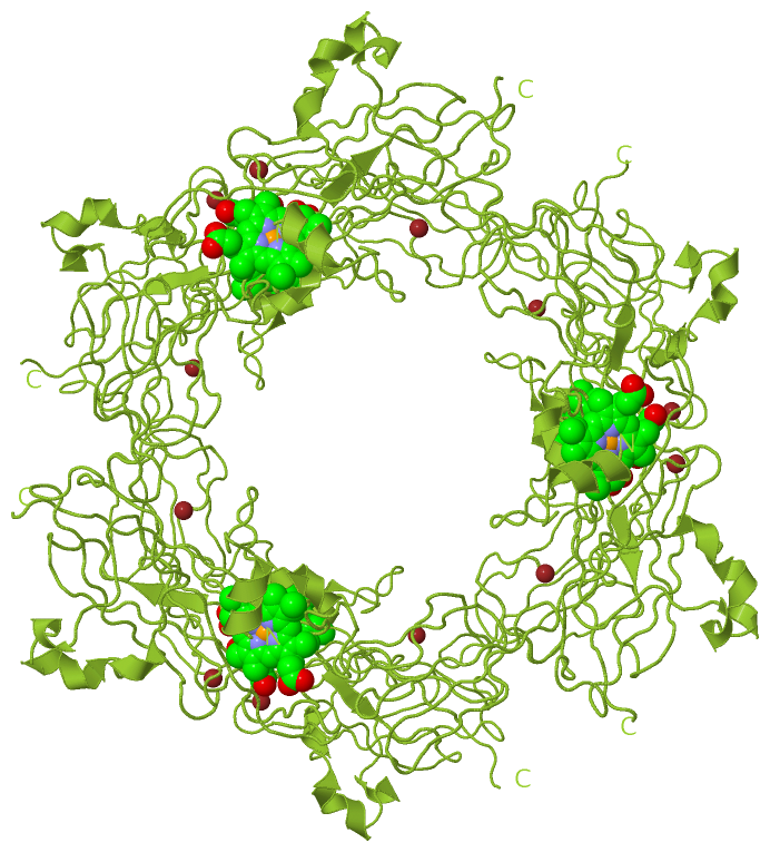
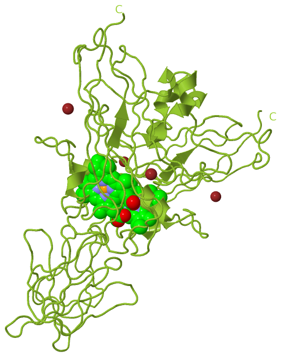
Ligands, Modified Residues, Ions (2, 3)| Asymmetric Unit (2, 3) Biological Unit 1 (1, 1) Biological Unit 2 (1, 6) Biological Unit 3 (1, 2) |
Sites (3, 3)
Asymmetric Unit (3, 3)
|
SS Bonds (0, 0)| (no "SS Bond" information available for 1CI3) |
Cis Peptide Bonds (1, 1)
Asymmetric Unit
|
||||||||
SAPs(SNPs)/Variants (0, 0)| (no "SAP(SNP)/Variant" information available for 1CI3) |
PROSITE Motifs (0, 0)| (no "PROSITE Motif" information available for 1CI3) |
Exons (0, 0)| (no "Exon" information available for 1CI3) |
Sequences/Alignments
Asymmetric UnitChain M from PDB Type:PROTEIN Length:249 aligned with CYF_PHOLA | P95522 from UniProtKB/Swiss-Prot Length:338 Alignment length:249 55 65 75 85 95 105 115 125 135 145 155 165 175 185 195 205 215 225 235 245 255 265 275 285 CYF_PHOLA 46 YPFWAQQNYANPREATGRIVCANCHLAAKPAEIEVPQAVLPDSVFKAVVKIPYDHSVQQVQADGSKGPLNVGAVLMLPEGFTIAPEDRIPEEMKEEVGPSYLFQPYADDKQNIVLVGPLPGDQYEEIVFPVLSPNPATNKSVAFGKYSIHLGANRGRGQIYPTGEKSNNAVYNASAAGVITAIAKADDGSAEVKIRTEDGTTIVDKIPAGPELIVSEGEEVAAGAALTNNPNVGGFGQKDTEIVLQSPN 294 SCOP domains d1ci3m1 M:1-169,M:232-249 Cytochrome f, large domain d1ci3m2 M:170-231 Cytochrome f, small domain d1ci3m1 SCOP domains CATH domains 1ci3M01 M:1-171,M:230-248 [code=2.60.40.830, no name defined] 1ci3M02 M:172-229 [code=2.40.50.100, no name defined] 1ci3M01 - CATH domains Pfam domains --------------------------------------------------------------------------------------------------------------------------------------------------------------------------------------------------------------------------------------------------------- Pfam domains SAPs(SNPs) --------------------------------------------------------------------------------------------------------------------------------------------------------------------------------------------------------------------------------------------------------- SAPs(SNPs) PROSITE --------------------------------------------------------------------------------------------------------------------------------------------------------------------------------------------------------------------------------------------------------- PROSITE Transcript --------------------------------------------------------------------------------------------------------------------------------------------------------------------------------------------------------------------------------------------------------- Transcript 1ci3 M 1 YPFWAQQNYANPREATGRIVCANCHLAAKPAEIEVPQAVLPDSVFKAVVKIPYDHSVQQVQADGSKGPLNVGAVLMLPEGFTIAPEDRIPEEMKEEVGPSYLFQPYADDKQNIVLVGPLPGDEYEEIVFPVLSPNPATNKSVAFGKYSIHLGANRGRGQIYPTGEKSNNAVYNASAAGVITAIAKADDGSAEVKIRTEDGTTIVDKIPAGPELIVSEGEEVAAGAALTNNPNVGGFGQKDTEIVLQSPN 249 10 20 30 40 50 60 70 80 90 100 110 120 130 140 150 160 170 180 190 200 210 220 230 240
|
||||||||||||||||||||
SCOP Domains (2, 2)
Asymmetric Unit
 |
CATH Domains (2, 2)
Asymmetric Unit
|
Pfam Domains (0, 0)| (no "Pfam Domain" information available for 1CI3) |
Gene Ontology (11, 11)|
Asymmetric Unit(hide GO term definitions) Chain M (CYF_PHOLA | P95522)
|
||||||||||||||||||||||||||||||||||||||||||||||||||||||||||||||||||||||||||||||||||||
Interactive Views
|
||||||||||||||||||||||||||||||||||||||||||||||||||||||||||||||||||||||||||||||||||||||||||||||||||||||||||||||||||||||||||||||||||||||||||||||||||||||||||||||||||||||||
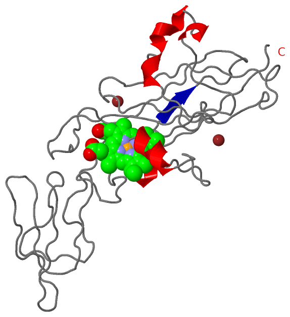
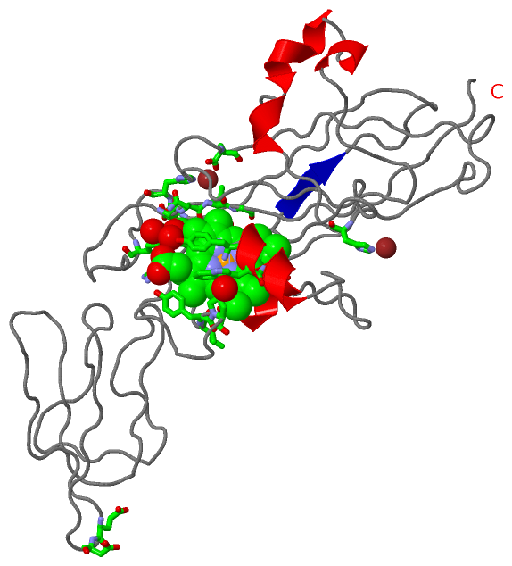
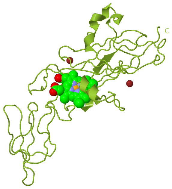
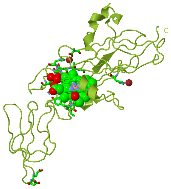
Still Images
|
||||||||||||||||
Databases
|
||||||||||||||||||||||||||||||||||||||||||||||||||||||||||||||||||||||||||||||||||||||||||||||||||||||||||||||||||||||||||||||||||||||||||||||||||||||||||||||||
Analysis Tools
|
|||||||||||||||||||||||||||||||||||||||||||||||||||||||||||||
Entries Sharing at Least One Protein Chain (UniProt ID)
Related Entries Specified in the PDB File
|
|