|
|
|
|
 Description
|
|
Compounds
|
||||||||||||||||||||||||||||||||||||||||
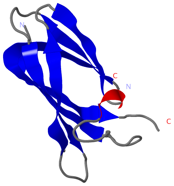
Chains, Units
Summary Information (see also Sequences/Alignments below) |
Ligands, Modified Residues, Ions (0, 0)| (no "Ligand,Modified Residues,Ions" information available for 2IIA) |
Sites (0, 0)| (no "Site" information available for 2IIA) |
SS Bonds (0, 0)| (no "SS Bond" information available for 2IIA) |
Cis Peptide Bonds (2, 2)
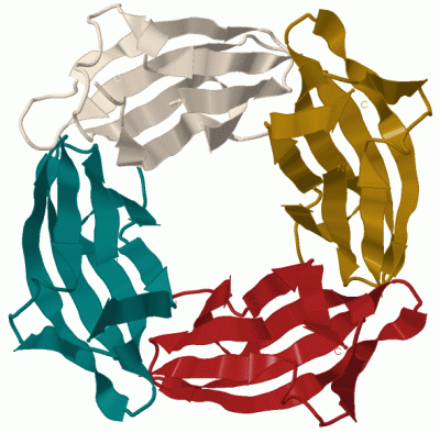
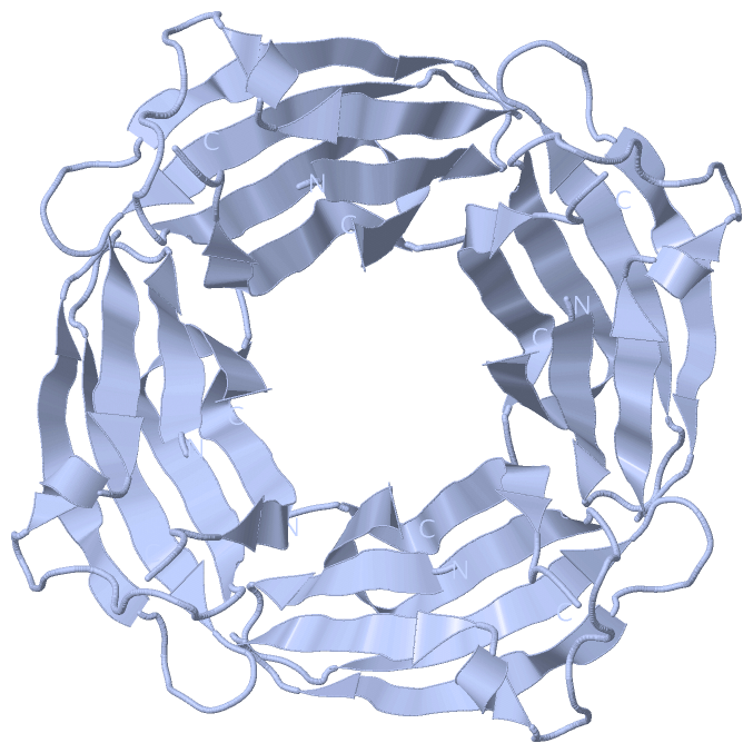
Asymmetric Unit
|
||||||||||||
SAPs(SNPs)/Variants (0, 0)| (no "SAP(SNP)/Variant" information available for 2IIA) |
PROSITE Motifs (0, 0)| (no "PROSITE Motif" information available for 2IIA) |
Exons (0, 0)| (no "Exon" information available for 2IIA) |
Sequences/Alignments
Asymmetric UnitChain A from PDB Type:PROTEIN Length:89 aligned with Q8YSC3_NOSS1 | Q8YSC3 from UniProtKB/TrEMBL Length:125 Alignment length:101 14 24 34 44 54 64 74 84 94 104 Q8YSC3_NOSS1 5 IGRTCWAIAEGYIPPYGNGPEPQFISHETVCILNAGDEDAHVEITIYYSDKEPVGPYRLTVPARRTKHVRFNDLNDPAPIPHDTDFASVIQSNVPIVVQHT 105 SCOP domains d2iiaa_ A: auto mated matches SCOP domains CATH domains ----------------------------------------------------------------------------------------------------- CATH domains Pfam domains ----------------------------------------------------------------------------------------------------- Pfam domains SAPs(SNPs) ----------------------------------------------------------------------------------------------------- SAPs(SNPs) PROSITE ----------------------------------------------------------------------------------------------------- PROSITE Transcript ----------------------------------------------------------------------------------------------------- Transcript 2iia A 4 IGRTCWAIAEGYIPP------------ETVCILNAGDEDAHVEITIYYSDKEPVGPYRLTVPARRTKHVRFNDLNDPAPIPHDTDFASVIQSNVPIVVQHT 104 13 | - |33 43 53 63 73 83 93 103 18 31
|
||||||||||||||||||||
SCOP Domains (1, 1)
Asymmetric Unit
 |
CATH Domains (0, 0)| (no "CATH Domain" information available for 2IIA) |
Pfam Domains (0, 0)| (no "Pfam Domain" information available for 2IIA) |
Gene Ontology (0, 0)|
Asymmetric Unit(hide GO term definitions)
(no "Gene Ontology" information available for 2IIA)
|
Interactive Views
|
||||||||||||||||||||||||||||||||||||||||||||||||||||||||||||||||||||||||||||||||||||||||||||||||||||||||||||||||||||||||||||||||||||||||||||||
Still Images
|
||||||||||||||||
Databases
|
||||||||||||||||||||||||||||||||||||||||||||||||||||||||||||||||||||||||||||||||||||||||||||||||||||||||||||||||||||||||||||||||||||||||||||||||||||||||||||||||
Analysis Tools
|
|||||||||||||||||||||||||||||||||||||||||||||||||||||||||||||
Entries Sharing at Least One Protein Chain (UniProt ID)
Related Entries Specified in the PDB File
|
|