|
|
|
|
 Description
|
|
Compounds
|
||||||||||||||||||||||||||||||||||||||||||||
Chains, Units
Summary Information (see also Sequences/Alignments below) |
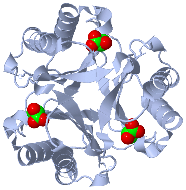
Ligands, Modified Residues, Ions (1, 1)
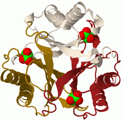
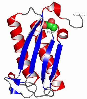
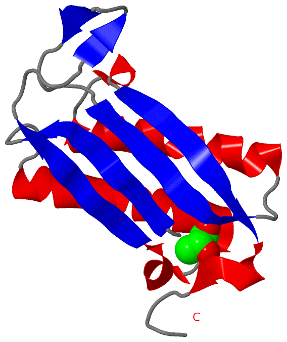
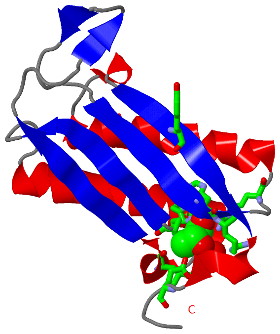
Asymmetric Unit (1, 1)
|
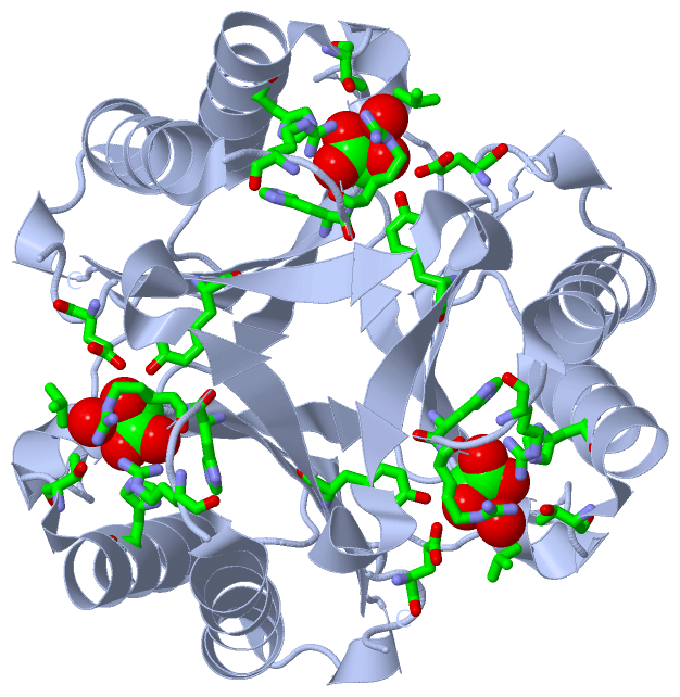
Sites (1, 1)
Asymmetric Unit (1, 1)
|
SS Bonds (0, 0)| (no "SS Bond" information available for 2FLT) |
Cis Peptide Bonds (0, 0)| (no "Cis Peptide Bond" information available for 2FLT) |
SAPs(SNPs)/Variants (0, 0)| (no "SAP(SNP)/Variant" information available for 2FLT) |
PROSITE Motifs (0, 0)| (no "PROSITE Motif" information available for 2FLT) |
Exons (0, 0)| (no "Exon" information available for 2FLT) |
Sequences/Alignments
Asymmetric UnitChain A from PDB Type:PROTEIN Length:117 aligned with Q6VPE5_9CORY | Q6VPE5 from UniProtKB/TrEMBL Length:150 Alignment length:117 11 21 31 41 51 61 71 81 91 101 111 Q6VPE5_9CORY 2 PVYMVYVSQDRLTPSAKHAVAKAITDAHRGLTGTQHFLAQVNFQEQPAGNVFLGGVQQGGDTIFVHGLHREGRSADLKGQLAQRIVDDVSVAAEIDRKHIWVYFGEMPAQQMVEYGR 118 SCOP domains --------------------------------------------------------------------------------------------------------------------- SCOP domains CATH domains --------------------------------------------------------------------------------------------------------------------- CATH domains Pfam domains --------------------------------------------------------------------------------------------------------------------- Pfam domains SAPs(SNPs) --------------------------------------------------------------------------------------------------------------------- SAPs(SNPs) PROSITE --------------------------------------------------------------------------------------------------------------------- PROSITE Transcript --------------------------------------------------------------------------------------------------------------------- Transcript 2flt A 1 PVYMVYVSQDRLTPSAKHAVAKAITDAHRGLTGTQHFLAQVNFNEQPAGNVFLGGVQQGGDTIFVHGLHREGRSADLKGQLAQRIVDDVSVAAEIDRKHIWVYFGEMPAQQMVEYGR 117 10 20 30 40 50 60 70 80 90 100 110
|
||||||||||||||||||||
SCOP Domains (0, 0)| (no "SCOP Domain" information available for 2FLT) |
CATH Domains (0, 0)| (no "CATH Domain" information available for 2FLT) |
Pfam Domains (0, 0)| (no "Pfam Domain" information available for 2FLT) |
Gene Ontology (0, 0)|
Asymmetric Unit(hide GO term definitions)
(no "Gene Ontology" information available for 2FLT)
|
Interactive Views
|
||||||||||||||||||||||||||||||||||||||||||||||||||||||||||||||||||||||||||||||||||||||||||||||||||||||||||||||||||||||||||||||||||||||||
Still Images
|
||||||||||||||||
Databases
|
||||||||||||||||||||||||||||||||||||||||||||||||||||||||||||||||||||||||||||||||||||||||||||||||||||||||||||||||||||||||||||||||||||||||||||||||||||||||||||||||
Analysis Tools
|
|||||||||||||||||||||||||||||||||||||||||||||||||||||||||||||
Entries Sharing at Least One Protein Chain (UniProt ID)
Related Entries Specified in the PDB File
|
|