Show PDB file:   
         Plain Text   HTML   (compressed file size)
QuickSearch:   
by PDB,NDB,UniProt,PROSITE Code or Search Term(s)  
(-)Asym./Biol. Unit
(-)Asym./Biol. Unit - sites
collapse expand < >
Image Asym./Biol. Unit
Asym./Biol. Unit  (Jmol Viewer)
Image Asym./Biol. Unit - sites
Asym./Biol. Unit - sites  (Jmol Viewer)

(-) Description

Title :  STRUCTURE OF THE ARTT MOTIF Q212A MUTANT C3BOT1 EXOENZYME ( FREE STATE, CRYSTAL FORM II)
 
Authors :  E. A. Stura, J. Menetrey, G. Flatau, P. Boquet, A. Menez
Date :  03 Dec 05  (Deposition) - 27 Feb 07  (Release) - 08 Jun 11  (Revision)
Method :  X-RAY DIFFRACTION
Resolution :  1.70
Chains :  Asym./Biol. Unit :  X
Keywords :  C3 Exoenzyme, Artt Motif, Bacterial Toxins, Transferase, Glycosyltransferase (Keyword Search: [Gene Ontology, PubMed, Web (Google))
 
Reference :  J. Menetrey, G. Flatau, P. Boquet, A. Menez, E. A. Stura
Structural Basis For The Nad-Hydrolysis Mechanism And The Artt-Loop Plasticity Of C3 Exoenzymes.
Protein Sci. V. 17 878 2008
PubMed-ID: 18369192  |  Reference-DOI: 10.1110/PS.073398508
(for further references see the PDB file header)

(-) Compounds

Molecule 1 - MONO-ADP-RIBOSYLTRANSFERASE C3
    ChainsX
    EC Number2.4.2.-
    EngineeredYES
    Expression SystemESCHERICHIA COLI
    Expression System StrainBL21
    Expression System Taxid511693
    MutationYES
    Organism ScientificCLOSTRIDIUM BOTULINUM D BACTERIOPHAGE
    Organism Taxid29342
    SynonymEXOENZYME C3

 Structural Features

(-) Chains, Units

  1
Asymmetric/Biological Unit X

Summary Information (see also Sequences/Alignments below)

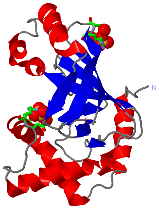
(-) Ligands, Modified Residues, Ions  (1, 2)

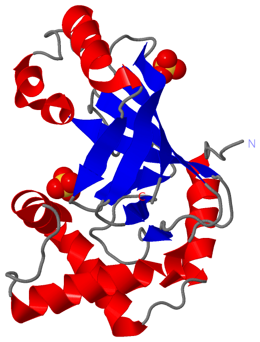
Asymmetric/Biological Unit (1, 2)
No.NameCountTypeFull Name
1SO42Ligand/IonSULFATE ION

(-) Sites  (2, 2)

Asymmetric Unit (2, 2)
No.NameEvidenceResiduesDescription
1AC1SOFTWAREARG X:229 , SER X:232 , HOH X:2227 , HOH X:2228BINDING SITE FOR RESIDUE SO4 X1246
2AC2SOFTWARESER X:84 , ASN X:87 , ARG X:91 , HOH X:2229BINDING SITE FOR RESIDUE SO4 X1247

(-) SS Bonds  (0, 0)

(no "SS Bond" information available for 2C8B)

(-) Cis Peptide Bonds  (0, 0)

(no "Cis Peptide Bond" information available for 2C8B)

 Sequence-Structure Mapping

(-) SAPs(SNPs)/Variants  (0, 0)

(no "SAP(SNP)/Variant" information available for 2C8B)

(-) PROSITE Motifs  (0, 0)

(no "PROSITE Motif" information available for 2C8B)

(-) Exons   (0, 0)

(no "Exon" information available for 2C8B)

(-) Sequences/Alignments

Asymmetric/Biological Unit
   Reformat: Number of residues per line =  ('0' or empty: single-line sequence representation)
  Number of residues per labelling interval =   
  UniProt sequence: complete  aligned part    
   Show mapping: SCOP domains CATH domains Pfam domains Secondary structure (by author)
SAPs(SNPs) PROSITE motifs Exons
(details for a mapped element are shown in a popup box when the mouse pointer rests over it)
Chain X from PDB  Type:PROTEIN  Length:201
 aligned with ARC3_CBDP | P15879 from UniProtKB/Swiss-Prot  Length:251

    Alignment length:201
                                    54        64        74        84        94       104       114       124       134       144       154       164       174       184       194       204       214       224       234       244 
            ARC3_CBDP    45 TYQEFTNIDQAKAWGNAQYKKYGLSKSEKEAIVSYTKSASEINGKLRQNKGVINGFPSNLIKQVELLDKSFNKMKTPENIMLFRGDDPAYLGTEFQNTLLNSNGTINKTAFEKAKAKFLNKDRLEYGYISTSLMNVSQFAGRPIITKFKVAKGSKAGYIDPISAFAGQLEMLLPRHSTYHIDDMRLSSDGKQIIITATMMG 245
               SCOP domains d2c8bx_ X: automated matches                                                                                                                                                                              SCOP domains
               CATH domains 2c8bX00 X:45-245 Toxin ADP-ribosyltransferase; Chain A, domain 1                                                                                                                                          CATH domains
               Pfam domains --------------------------------------------------------------------------------------------------------------------------------------------------------------------------------------------------------- Pfam domains
         Sec.struct. author ......hhhhhhhhhhhhhh....hhhhhhhhhhhhhhhhhhhhhhhhh.......hhhhhhhhhhhhhhhhhh.....eeeeeeehhhhhhhhhhh.........hhhhhhhhhhhhh..eeee...eeee...........eeeeeee......ee...........eeee...eeeeeeeeee......eeeeeee.. Sec.struct. author
                 SAPs(SNPs) --------------------------------------------------------------------------------------------------------------------------------------------------------------------------------------------------------- SAPs(SNPs)
                    PROSITE --------------------------------------------------------------------------------------------------------------------------------------------------------------------------------------------------------- PROSITE
                 Transcript --------------------------------------------------------------------------------------------------------------------------------------------------------------------------------------------------------- Transcript
                 2c8b X  45 TYQEFTNIDQAKAWGNAQYKKYGLSKSEKEAIVSYTKSASEINGKLRQNKGVINGFPSNLIKQVELLDKSFNKMKTPENIMLFRGDDPAYLGTEFQNTLLNSNGTINKTAFEKAKAKFLNKDRLEYGYISTSLMNVSQFAGRPIITKFKVAKGSKAGYIDPISAFAGALEMLLPRHSTYHIDDMRLSSDGKQIIITATMMG 245
                                    54        64        74        84        94       104       114       124       134       144       154       164       174       184       194       204       214       224       234       244 

   Legend:   → Mismatch (orange background)
  - → Gap (green background, '-', border residues have a numbering label)
    → Modified Residue (blue background, lower-case, 'x' indicates undefined single-letter code, labelled with number + name)
  x → Chemical Group (purple background, 'x', labelled with number + name, e.g. ACE or NH2)
  extra numbering lines below/above indicate numbering irregularities and modified residue names etc., number ends below/above '|'

 Classification and Annotation

(-) SCOP Domains  (1, 1)

Asymmetric/Biological Unit

(-) CATH Domains  (1, 1)

Asymmetric/Biological Unit
(-)
Class: Alpha Beta (26913)

(-) Pfam Domains  (0, 0)

(no "Pfam Domain" information available for 2C8B)

(-) Gene Ontology  (7, 7)

Asymmetric/Biological Unit(hide GO term definitions)
Chain X   (ARC3_CBDP | P15879)
molecular function
    GO:0016740    transferase activity    Catalysis of the transfer of a group, e.g. a methyl group, glycosyl group, acyl group, phosphorus-containing, or other groups, from one compound (generally regarded as the donor) to another compound (generally regarded as the acceptor). Transferase is the systematic name for any enzyme of EC class 2.
    GO:0016757    transferase activity, transferring glycosyl groups    Catalysis of the transfer of a glycosyl group from one compound (donor) to another (acceptor).
    GO:0016763    transferase activity, transferring pentosyl groups    Catalysis of the transfer of a pentosyl group from one compound (donor) to another (acceptor).
biological process
    GO:0009405    pathogenesis    The set of specific processes that generate the ability of an organism to induce an abnormal, generally detrimental state in another organism.
    GO:0006471    protein ADP-ribosylation    The transfer, from NAD, of ADP-ribose to protein amino acids.
    GO:0016032    viral process    A multi-organism process in which a virus is a participant. The other participant is the host. Includes infection of a host cell, replication of the viral genome, and assembly of progeny virus particles. In some cases the viral genetic material may integrate into the host genome and only subsequently, under particular circumstances, 'complete' its life cycle.
cellular component
    GO:0005576    extracellular region    The space external to the outermost structure of a cell. For cells without external protective or external encapsulating structures this refers to space outside of the plasma membrane. This term covers the host cell environment outside an intracellular parasite.

 Visualization

(-) Interactive Views

Asymmetric/Biological Unit
  Complete Structure
    Jena3D(integrated viewing of ligand, site, SAP, PROSITE, SCOP information)
    WebMol | AstexViewer[tm]@PDBe
(Java Applets, require no local installation except for Java; loading may be slow)
    STRAP
(Java WebStart application, automatic local installation, requires Java; full application with system access!)
    RasMol
(require local installation)
    Molscript (VRML)
(requires installation of a VRML viewer; select preferred view via VRML and generate a mono or stereo PDF format file)
 
  Ligands, Modified Residues, Ions
    SO4  [ RasMol | Jena3D ]  +environment [ RasMol | Jena3D ]
 
  Sites
    AC1  [ RasMol ]  +environment [ RasMol ]
    AC2  [ RasMol ]  +environment [ RasMol ]
 
  Cis Peptide Bonds
(no "Cis Peptide Bonds" information available for 2c8b)
 

(-) Still Images

Jmol
  protein: cartoon or spacefill or dots and stick; nucleic acid: cartoon and stick; ligands: spacefill; active site: stick
Molscript
  protein, nucleic acid: cartoon; ligands: spacefill; active site: ball and stick

 Databases and Analysis Tools

(-) Databases

Access by PDB/NDB ID
  2c8b
    Family and Domain InformationProDom | SYSTERS
    General Structural InformationGlycoscienceDB | MMDB | NDB | OCA | PDB | PDBe | PDBj | PDBsum | PDBWiki | PQS | PROTEOPEDIA
    Orientation in MembranesOPM
    Protein SurfaceSURFACE
    Secondary StructureDSSP (structure derived) | HSSP (homology derived)
    Structural GenomicsGeneCensus
    Structural NeighboursCE | VAST
    Structure ClassificationCATH | Dali | SCOP
    Validation and Original DataBMRB Data View | BMRB Restraints Grid | EDS | PROCHECK | RECOORD | WHAT_CHECK
 
Access by UniProt ID/Accession number
  ARC3_CBDP | P15879
    Comparative Protein Structure ModelsModBase
    Genomic InformationEnsembl
    Protein-protein InteractionDIP
    Sequence, Family and Domain InformationInterPro | Pfam | SMART | UniProtKB/SwissProt
 
Access by Enzyme Classificator   (EC Number)
  2.4.2.-
    General Enzyme InformationBRENDA | EC-PDB | Enzyme | IntEnz
    PathwayKEGG | MetaCyc
 
Access by Disease Identifier   (MIM ID)
  (no 'MIM ID' available)
    Disease InformationOMIM
 
Access by GenAge ID
  (no 'GenAge ID' available)
    Age Related InformationGenAge

(-) Analysis Tools

Access by PDB/NDB ID
    Domain InformationXDom
    Interatomic Contacts of Structural UnitsCSU
    Ligand-protein ContactsLPC
    Protein CavitiescastP
    Sequence and Secondary StructurePDBCartoon
    Structure AlignmentSTRAP(Java WebStart application, automatic local installation, requires Java; full application with system access!)
    Structure and Sequence BrowserSTING
 
Access by UniProt ID/Accession number
  ARC3_CBDP | P15879
    Protein Disorder PredictionDisEMBL | FoldIndex | GLOBPLOT (for more information see DisProt)

 Related Entries

(-) Entries Sharing at Least One Protein Chain (UniProt ID)

UniProtKB/Swiss-Prot
        ARC3_CBDP | P158791g24 1gze 1gzf 1uzi 2a78 2a9k 2bov 2c89 2c8a 2c8c 2c8d 2c8e 2c8f 2c8g 2c8h

(-) Related Entries Specified in the PDB File

1g24 THE CRYSTAL STRUCTURE OF EXOENZYME C3 FROM CLOSTRIDIUMBOTULINUM
1gze STRUCTURE OF THE CLOSTRIDIUM BOTULINUM C3 EXOENZYME (L177C MUTANT)
1gzf STRUCTURE OF THE CLOSTRIDIUM BOTULINUM C3 EXOENZYME (WILD-TYPE) IN COMPLEX WITH NAD
1uzi C3 EXOENZYME FROM CLOSTRIDIUM BOTULINUM, TETRAGONAL FORM
2a78 CRYSTAL STRUCTURE OF THE C3BOT-RALA COMPLEX REVEALS A NOVELTYPE OF ACTION OF A BACTERIAL EXOENZYME
2a9k CRYSTAL STRUCTURE OF THE C3BOT-NAD-RALA COMPLEX REVEALS ANOVEL TYPE OF ACTION OF A BACTERIAL EXOENZYME
2bov MOLECULAR RECOGNITION OF AN ADP-RIBOSYLATING CLOSTRIDIUM BOTULINUM C3 EXOENZYME BY RALA GTPASE
2c89 STRUCTURE OF THE WILD-TYPE C3BOT1 EXOENZYME (FREE STATE, CRYSTAL FORM I)
2c8a STRUCTURE OF THE WILD-TYPE C3BOT1 EXOENZYME (NICOTINAMIDE-BOUND STATE, CRYSTAL FORM I)
2c8c STRUCTURE OF THE ARTT MOTIF Q212A MUTANT C3BOT1 EXOENZYME (NAD-BOUND STATE, CRYSTAL FORM I
2c8d STRUCTURE OF THE ARTT MOTIF Q212A MUTANT C3BOT1 EXOENZYME (FREE STATE, CRYSTAL FORM I)
2c8e STRUCTURE OF THE ARTT MOTIF E214N MUTANT C3BOT1 EXOENZYME (FREE STATE, CRYSTAL FORM III)
2c8f STRUCTURE OF THE ARTT MOTIF E214N MUTANT C3BOT1 EXOENZYME (NAD-BOUND STATE, CRYSTAL FORM III)
2c8g STRUCTURE OF THE PN LOOP Q182A MUTANT C3BOT1 EXOENZYME (FREE STATE, CRYSTAL FORM I)
2c8h STRUCTURE OF THE PN LOOP Q182A MUTANT C3BOT1 EXOENZYME (NAD-BOUND STATE, CRYSTAL FORM I)