Show PDB file:   
         Plain Text   HTML   (compressed file size)
QuickSearch:   
by PDB,NDB,UniProt,PROSITE Code or Search Term(s)  
(-)Asym.Unit - manually
(-)Asymmetric Unit
(-)Asym. Unit - sites
(-)Biological Unit 1
(-)Biol. Unit 1 - sites
(-)Biological Unit 2
(-)Biological Unit 3
(-)Biological Unit 4
collapse expand < >
Image Asym.Unit - manually
Asym.Unit - manually  (Jmol Viewer)
Image Asymmetric Unit
Asymmetric Unit  (Jmol Viewer)
Image Asym. Unit - sites
Asym. Unit - sites  (Jmol Viewer)
Image Biological Unit 1
Biological Unit 1  (Jmol Viewer)
Image Biol. Unit 1 - sites
Biol. Unit 1 - sites  (Jmol Viewer)
Image Biological Unit 2
Biological Unit 2  (Jmol Viewer)
Image Biological Unit 3
Biological Unit 3  (Jmol Viewer)
Image Biological Unit 4
Biological Unit 4  (Jmol Viewer)

(-) Description

Title :  STRUCTURE OF A NOVEL PHOTORECEPTOR: THE BLUF DOMAIN OF APPA FROM RHODOBACTER SPHAEROIDES
 
Authors :  S. Anderson, V. Dragnea, S. Masuda, J. Ybe, K. Moffat, C. Bauer
Date :  05 Feb 05  (Deposition) - 28 Jun 05  (Release) - 13 Jul 11  (Revision)
Method :  X-RAY DIFFRACTION
Resolution :  2.30
Chains :  Asym. Unit :  A,B,C
Biol. Unit 1:  A,B  (1x)
Biol. Unit 2:  C  (2x)
Biol. Unit 3:  A (1x),B (1x),C (2x)
Biol. Unit 4:  A,B,C  (2x)
Keywords :  Ferredoxin-Like Fold, Flavin Binding, Photoreceptor, Transcription (Keyword Search: [Gene Ontology, PubMed, Web (Google))
 
Reference :  S. Anderson, V. Dragnea, S. Masuda, J. Ybe, K. Moffat, C. Bauer
Structure Of A Novel Photoreceptor, The Bluf Domain Of Appa From Rhodobacter Sphaeroides
Biochemistry V. 44 7998 2005
PubMed-ID: 15924418  |  Reference-DOI: 10.1021/BI0502691

(-) Compounds

Molecule 1 - HYPOTHETICAL PROTEIN RSPH03001874
    ChainsA, B, C
    EngineeredYES
    Expression SystemESCHERICHIA COLI
    Expression System Taxid562
    FragmentSEQUENCE DATABASE RESIDUES 17-133
    Organism ScientificRHODOBACTER SPHAEROIDES 2.4.1
    Organism Taxid272943
    StrainATCC 17023 / 2.4.1 / NCIB 8253 / DSM 158
    SynonymAPPA BLUF DOMAIN

 Structural Features

(-) Chains, Units

  123
Asymmetric Unit ABC
Biological Unit 1 (1x)AB 
Biological Unit 2 (2x)  C
Biological Unit 3 (2x)A (1x)B (1x)C (2x)
Biological Unit 4 (2x)ABC

Summary Information (see also Sequences/Alignments below)

(-) Ligands, Modified Residues, Ions  (2, 9)

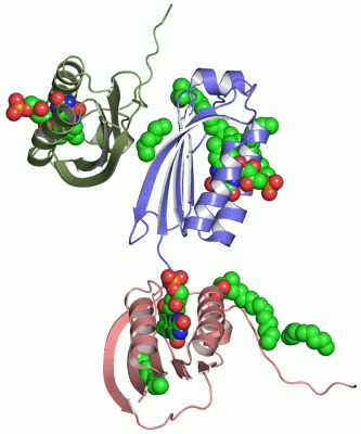
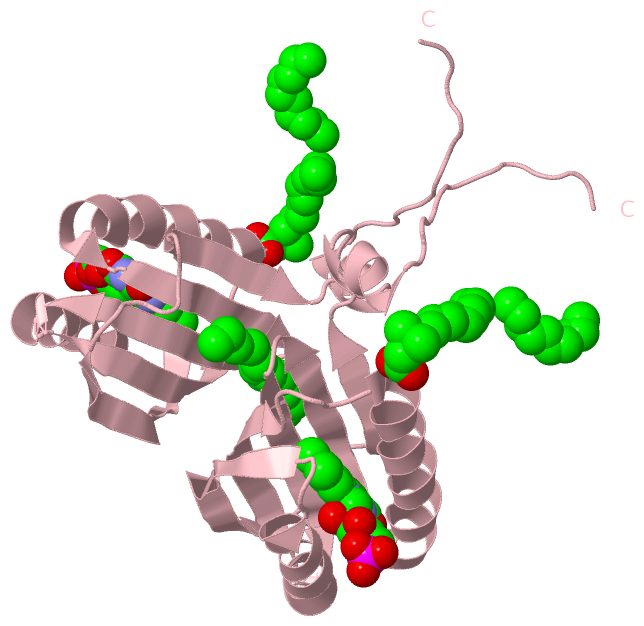
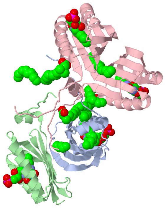
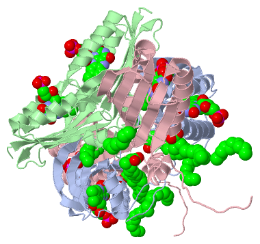
Asymmetric Unit (2, 9)
No.NameCountTypeFull Name
1D9G6Ligand/IonN-DODECYL-N,N-DIMETHYLGLYCINATE
2FMN3Ligand/IonFLAVIN MONONUCLEOTIDE
Biological Unit 1 (2, 5)
No.NameCountTypeFull Name
1D9G3Ligand/IonN-DODECYL-N,N-DIMETHYLGLYCINATE
2FMN2Ligand/IonFLAVIN MONONUCLEOTIDE
Biological Unit 2 (2, 8)
No.NameCountTypeFull Name
1D9G6Ligand/IonN-DODECYL-N,N-DIMETHYLGLYCINATE
2FMN2Ligand/IonFLAVIN MONONUCLEOTIDE
Biological Unit 3 (0, 0)
No.NameCountTypeFull Name
1D9G-1Ligand/IonN-DODECYL-N,N-DIMETHYLGLYCINATE
2FMN-1Ligand/IonFLAVIN MONONUCLEOTIDE
Biological Unit 4 (2, 18)
No.NameCountTypeFull Name
1D9G12Ligand/IonN-DODECYL-N,N-DIMETHYLGLYCINATE
2FMN6Ligand/IonFLAVIN MONONUCLEOTIDE

(-) Sites  (8, 8)

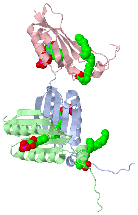
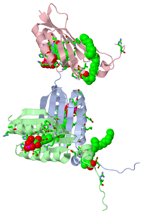
Asymmetric Unit (8, 8)
No.NameEvidenceResiduesDescription
1AC1SOFTWARETYR A:21 , ILE A:37 , THR A:40 , SER A:41 , HIS A:44 , ASN A:45 , ARG A:47 , PHE A:61 , GLN A:63 , VAL A:75 , HIS A:78 , ILE A:79 , ASP A:82 , ARG A:84 , HIS A:85 , HOH A:343 , HOH A:351BINDING SITE FOR RESIDUE FMN A 201
2AC2SOFTWARETYR B:21 , THR B:40 , SER B:41 , HIS B:44 , ASN B:45 , PHE B:61 , GLN B:63 , VAL B:75 , HIS B:78 , ILE B:79 , ASP B:82 , ARG B:84 , HIS B:85 , HOH B:222BINDING SITE FOR RESIDUE FMN B 202
3AC3SOFTWAREARG B:68 , ALA B:70 , TYR C:21 , ILE C:37 , THR C:40 , SER C:41 , HIS C:44 , ASN C:45 , PHE C:61 , GLN C:63 , VAL C:75 , HIS C:78 , ILE C:79 , ARG C:81 , ASP C:82 , ARG C:84 , HIS C:85 , HOH C:324BINDING SITE FOR RESIDUE FMN C 203
4AC4SOFTWARELEU A:35 , GLU A:39 , LEU A:108 , MET A:115 , THR C:30 , LEU C:31 , ARG C:32 , LEU C:118 , D9G C:305BINDING SITE FOR RESIDUE D9G C 301
5AC5SOFTWAREGLU A:122 , ARG B:124 , ARG C:124BINDING SITE FOR RESIDUE D9G A 302
6AC6SOFTWAREGLN C:58 , PHE C:62BINDING SITE FOR RESIDUE D9G C 304
7AC7SOFTWARELEU A:34 , TYR A:56 , CYS A:110 , ASP A:114 , LEU A:118 , D9G C:301BINDING SITE FOR RESIDUE D9G C 305
8AC8SOFTWAREPHE A:62 , PHE B:55BINDING SITE FOR RESIDUE D9G A 306

(-) SS Bonds  (0, 0)

(no "SS Bond" information available for 1YRX)

(-) Cis Peptide Bonds  (0, 0)

(no "Cis Peptide Bond" information available for 1YRX)

 Sequence-Structure Mapping

(-) SAPs(SNPs)/Variants  (0, 0)

(no "SAP(SNP)/Variant" information available for 1YRX)

(-) PROSITE Motifs  (0, 0)

(no "PROSITE Motif" information available for 1YRX)

(-) Exons   (0, 0)

(no "Exon" information available for 1YRX)

(-) Sequences/Alignments

Asymmetric Unit
   Reformat: Number of residues per line =  ('0' or empty: single-line sequence representation)
  Number of residues per labelling interval =   
  UniProt sequence: complete  aligned part    
   Show mapping: SCOP domains CATH domains Pfam domains Secondary structure (by author)
SAPs(SNPs) PROSITE motifs Exons
(details for a mapped element are shown in a popup box when the mouse pointer rests over it)
Chain A from PDB  Type:PROTEIN  Length:118
 aligned with Q53119_RHOSH | Q53119 from UniProtKB/TrEMBL  Length:450

    Alignment length:119
                                    21        31        41        51        61        71        81        91       101       111       121         
         Q53119_RHOSH    12 TGSDLVSCCYRSLAAPDLTLRDLLDIVETSQAHNARAQLTGALFYSQGVFFQWLEGRPAAVAEVMTHIQRDRRHSNVEILAEEPIAKRRFAGWHMQLSCSEADMRSLGLAESRQIVTVG 130
               SCOP domains -----d1yrxa1 A:17-130 Sensor of blue light AppA                                                                         SCOP domains
               CATH domains ----------------------------------------------------------------------------------------------------------------------- CATH domains
               Pfam domains ----------------------------------------------------------------------------------------------------------------------- Pfam domains
         Sec.struct. author ..-.eeeeeeeeee....hhhhhhhhhhhhhhhhhhhh.eeeeeee..eeeeeeeehhhhhhhhhhhhhh...eeeeeeeeeeee.......eeeee..hhhhhhhh............ Sec.struct. author
                 SAPs(SNPs) ----------------------------------------------------------------------------------------------------------------------- SAPs(SNPs)
                    PROSITE ----------------------------------------------------------------------------------------------------------------------- PROSITE
                 Transcript ----------------------------------------------------------------------------------------------------------------------- Transcript
                 1yrx A  13 AG-HMVSCCYRSLAAPDLTLRDLLDIVETSQAHNARAQLTGALFYSQGVFFQWLEGRPAAVAEVMTHIQRDRRHSNVEILAEEPIAKRRFAGWHMQLSCSEADMRSLGLAESRQIVTVG 130
                             | |    21        31        41        51        61        71        81        91       101       111       121         
                             | |                                                                                                                   
                            14 |                                                                                                                   
                              15                                                                                                                   

Chain B from PDB  Type:PROTEIN  Length:116
 aligned with Q53119_RHOSH | Q53119 from UniProtKB/TrEMBL  Length:450

    Alignment length:116
                                    23        33        43        53        63        73        83        93       103       113       123      
         Q53119_RHOSH    14 SDLVSCCYRSLAAPDLTLRDLLDIVETSQAHNARAQLTGALFYSQGVFFQWLEGRPAAVAEVMTHIQRDRRHSNVEILAEEPIAKRRFAGWHMQLSCSEADMRSLGLAESRQIVTV 129
               SCOP domains d1yrxb_ B: automated matches                                                                                         SCOP domains
               CATH domains -------------------------------------------------------------------------------------------------------------------- CATH domains
               Pfam domains -------------------------------------------------------------------------------------------------------------------- Pfam domains
         Sec.struct. author ..eeeeeeeeee....hhhhhhhhhhhhhhhhh....eeeeeee..eeeeeeeehhhhhhhhhhhhhhh..eeeeeeeeeeee.......eeeee..hhhhhhh............ Sec.struct. author
                 SAPs(SNPs) -------------------------------------------------------------------------------------------------------------------- SAPs(SNPs)
                    PROSITE -------------------------------------------------------------------------------------------------------------------- PROSITE
                 Transcript -------------------------------------------------------------------------------------------------------------------- Transcript
                 1yrx B  14 GHMVSCCYRSLAAPDLTLRDLLDIVETSQAHNARAQLTGALFYSQGVFFQWLEGRPAAVAEVMTHIQRDRRHSNVEILAEEPIAKRRFAGWHMQLSCSEADMRSLGLAESRQIVTV 129
                                    23        33        43        53        63        73        83        93       103       113       123      

Chain C from PDB  Type:PROTEIN  Length:115
 aligned with Q53119_RHOSH | Q53119 from UniProtKB/TrEMBL  Length:450

    Alignment length:115
                                    24        34        44        54        64        74        84        94       104       114       124     
         Q53119_RHOSH    15 DLVSCCYRSLAAPDLTLRDLLDIVETSQAHNARAQLTGALFYSQGVFFQWLEGRPAAVAEVMTHIQRDRRHSNVEILAEEPIAKRRFAGWHMQLSCSEADMRSLGLAESRQIVTV 129
               SCOP domains d1yrxc_ C: automated matches                                                                                        SCOP domains
               CATH domains ------------------------------------------------------------------------------------------------------------------- CATH domains
           Pfam domains (1) --BLUF-1yrxC01 C:17-108                                                                       --------------------- Pfam domains (1)
           Pfam domains (2) --BLUF-1yrxC02 C:17-108                                                                       --------------------- Pfam domains (2)
           Pfam domains (3) --BLUF-1yrxC03 C:17-108                                                                       --------------------- Pfam domains (3)
         Sec.struct. author .eeeeeeeeee....hhhhhhhhhhhhhhhhhhhh.eeeeeee..eeeeeeeehhhhhhhhhhhhhh...eeeeeeeeeeee.......eeeee....hhhhhh........... Sec.struct. author
                 SAPs(SNPs) ------------------------------------------------------------------------------------------------------------------- SAPs(SNPs)
                    PROSITE ------------------------------------------------------------------------------------------------------------------- PROSITE
                 Transcript ------------------------------------------------------------------------------------------------------------------- Transcript
                 1yrx C  15 HMVSCCYRSLAAPDLTLRDLLDIVETSQAHNARAQLTGALFYSQGVFFQWLEGRPAAVAEVMTHIQRDRRHSNVEILAEEPIAKRRFAGWHMQLSCSEADMRSLGLAESRQIVTV 129
                                    24        34        44        54        64        74        84        94       104       114       124     

   Legend:   → Mismatch (orange background)
  - → Gap (green background, '-', border residues have a numbering label)
    → Modified Residue (blue background, lower-case, 'x' indicates undefined single-letter code, labelled with number + name)
  x → Chemical Group (purple background, 'x', labelled with number + name, e.g. ACE or NH2)
  extra numbering lines below/above indicate numbering irregularities and modified residue names etc., number ends below/above '|'

 Classification and Annotation

(-) SCOP Domains  (2, 3)

Asymmetric Unit

(-) CATH Domains  (0, 0)

(no "CATH Domain" information available for 1YRX)

(-) Pfam Domains  (1, 3)

Asymmetric Unit

(-) Gene Ontology  (4, 4)

Asymmetric Unit(hide GO term definitions)
Chain A,B,C   (Q53119_RHOSH | Q53119)
molecular function
    GO:0071949    FAD binding    Interacting selectively and non-covalently with the oxidized form, FAD, of flavin-adenine dinucleotide, the coenzyme or the prosthetic group of various flavoprotein oxidoreductase enzymes.
    GO:0009882    blue light photoreceptor activity    The function of absorbing and responding to electromagnetic radiation with a wavelength of approximately 400-470nm. The response may involve a change in conformation.
    GO:0000166    nucleotide binding    Interacting selectively and non-covalently with a nucleotide, any compound consisting of a nucleoside that is esterified with (ortho)phosphate or an oligophosphate at any hydroxyl group on the ribose or deoxyribose.
biological process
    GO:0009785    blue light signaling pathway    The series of molecular signals initiated upon sensing of blue light by photoreceptor molecule, at a wavelength between 400nm and 470nm.

 Visualization

(-) Interactive Views

Asymmetric Unit
  Complete Structure
    Jena3D(integrated viewing of ligand, site, SAP, PROSITE, SCOP information)
    WebMol | AstexViewer[tm]@PDBe
(Java Applets, require no local installation except for Java; loading may be slow)
    STRAP
(Java WebStart application, automatic local installation, requires Java; full application with system access!)
    RasMol
(require local installation)
    Molscript (VRML)
(requires installation of a VRML viewer; select preferred view via VRML and generate a mono or stereo PDF format file)
 
  Ligands, Modified Residues, Ions
    D9G  [ RasMol | Jena3D ]  +environment [ RasMol | Jena3D ]
    FMN  [ RasMol | Jena3D ]  +environment [ RasMol | Jena3D ]
 
  Sites
    AC1  [ RasMol ]  +environment [ RasMol ]
    AC2  [ RasMol ]  +environment [ RasMol ]
    AC3  [ RasMol ]  +environment [ RasMol ]
    AC4  [ RasMol ]  +environment [ RasMol ]
    AC5  [ RasMol ]  +environment [ RasMol ]
    AC6  [ RasMol ]  +environment [ RasMol ]
    AC7  [ RasMol ]  +environment [ RasMol ]
    AC8  [ RasMol ]  +environment [ RasMol ]
 
  Cis Peptide Bonds
(no "Cis Peptide Bonds" information available for 1yrx)
 
Biological Units
  Complete Structure
    Biological Unit 1  [ Jena3D ]
    Biological Unit 2  [ Jena3D ]
    Biological Unit 3  [ Jena3D ]
    Biological Unit 4  [ Jena3D ]

(-) Still Images

Jmol
  protein: cartoon or spacefill or dots and stick; nucleic acid: cartoon and stick; ligands: spacefill; active site: stick
Molscript
  protein, nucleic acid: cartoon; ligands: spacefill; active site: ball and stick

 Databases and Analysis Tools

(-) Databases

Access by PDB/NDB ID
  1yrx
    Family and Domain InformationProDom | SYSTERS
    General Structural InformationGlycoscienceDB | MMDB | NDB | OCA | PDB | PDBe | PDBj | PDBsum | PDBWiki | PQS | PROTEOPEDIA
    Orientation in MembranesOPM
    Protein SurfaceSURFACE
    Secondary StructureDSSP (structure derived) | HSSP (homology derived)
    Structural GenomicsGeneCensus
    Structural NeighboursCE | VAST
    Structure ClassificationCATH | Dali | SCOP
    Validation and Original DataBMRB Data View | BMRB Restraints Grid | EDS | PROCHECK | RECOORD | WHAT_CHECK
 
Access by UniProt ID/Accession number
  Q53119_RHOSH | Q53119
    Comparative Protein Structure ModelsModBase
    Genomic InformationEnsembl
    Protein-protein InteractionDIP
    Sequence, Family and Domain InformationInterPro | Pfam | SMART | UniProtKB/TrEMBL
 
Access by Enzyme Classificator   (EC Number)
  (no 'Enzyme Classificator' available)
    General Enzyme InformationBRENDA | EC-PDB | Enzyme | IntEnz
    PathwayKEGG | MetaCyc
 
Access by Disease Identifier   (MIM ID)
  (no 'MIM ID' available)
    Disease InformationOMIM
 
Access by GenAge ID
  (no 'GenAge ID' available)
    Age Related InformationGenAge

(-) Analysis Tools

Access by PDB/NDB ID
    Domain InformationXDom
    Interatomic Contacts of Structural UnitsCSU
    Ligand-protein ContactsLPC
    Protein CavitiescastP
    Sequence and Secondary StructurePDBCartoon
    Structure AlignmentSTRAP(Java WebStart application, automatic local installation, requires Java; full application with system access!)
    Structure and Sequence BrowserSTING
 
Access by UniProt ID/Accession number
  Q53119_RHOSH | Q53119
    Protein Disorder PredictionDisEMBL | FoldIndex | GLOBPLOT (for more information see DisProt)

 Related Entries

(-) Entries Sharing at Least One Protein Chain (UniProt ID)

UniProtKB/TrEMBL
        Q53119_RHOSH | Q531192bun 4heh 4hh0 4hh1 4hh3

(-) Related Entries Specified in the PDB File

(no "Related Entries Specified in the PDB File" available for 1YRX)