|
|
|
|
 Description
|
|
Compounds
|
||||||||||||||||||||||||
Chains, Units
Summary Information (see also Sequences/Alignments below) |
Ligands, Modified Residues, Ions (3, 11)
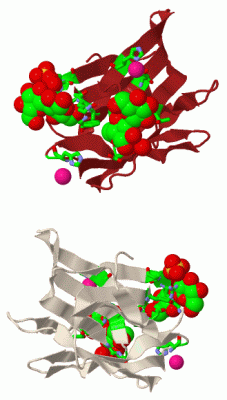
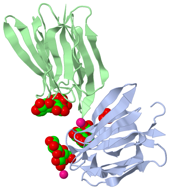
Asymmetric Unit (3, 11)
|
Sites (7, 7)
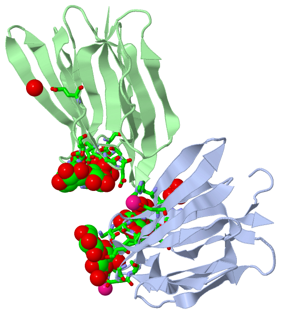
Asymmetric Unit (7, 7)
|
SS Bonds (0, 0)| (no "SS Bond" information available for 2BN0) |
Cis Peptide Bonds (2, 2)
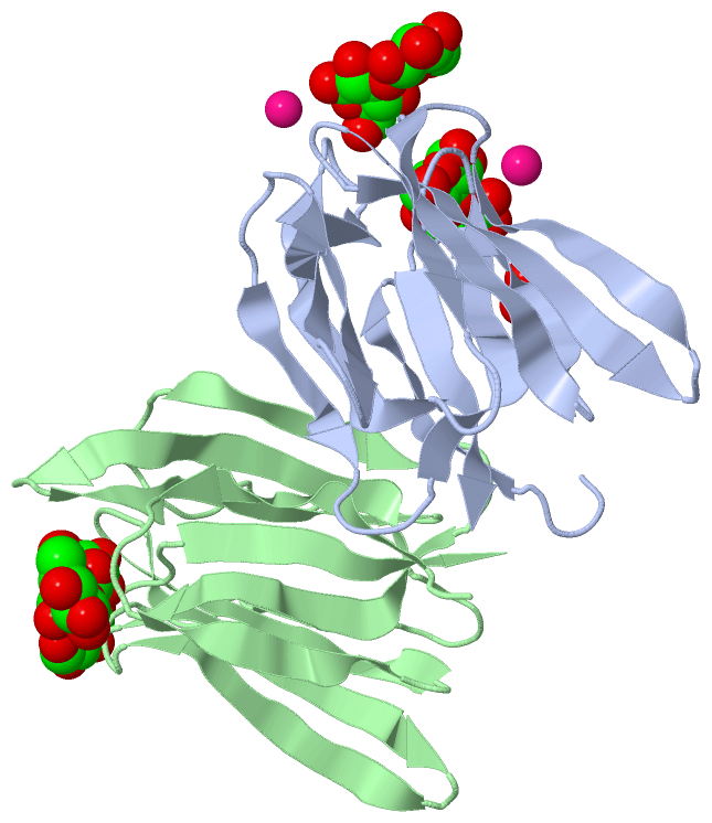
Asymmetric Unit
|
||||||||||||
SAPs(SNPs)/Variants (0, 0)| (no "SAP(SNP)/Variant" information available for 2BN0) |
PROSITE Motifs (0, 0)| (no "PROSITE Motif" information available for 2BN0) |
Exons (0, 0)| (no "Exon" information available for 2BN0) |
Sequences/Alignments
Asymmetric UnitChain A from PDB Type:PROTEIN Length:141 aligned with O22321_MUSAC | O22321 from UniProtKB/TrEMBL Length:141 Alignment length:141 10 20 30 40 50 60 70 80 90 100 110 120 130 140 O22321_MUSAC 1 MNGAIKVGAWGGNGGSAFDMGPAYRIISVKIFSGDVVDGVDVTFTYYGKTETRHYGGSGGTPHEIVLQEGEYLVGMAGEVANYHGAVVLGKLGFSTNKKAYGPFGNTGGTPFSLPIAAGKISGFFGRGGKFLDAIGVYLEP 141 SCOP domains d2bn0a_ A: automated matches SCOP domains CATH domains 2bn0A00 A:1-141 [code=2.100.10.30, no name defined] CATH domains Pfam domains --------------------------------------------------------------------------------------------------------------------------------------------- Pfam domains SAPs(SNPs) --------------------------------------------------------------------------------------------------------------------------------------------- SAPs(SNPs) PROSITE --------------------------------------------------------------------------------------------------------------------------------------------- PROSITE Transcript --------------------------------------------------------------------------------------------------------------------------------------------- Transcript 2bn0 A 1 MNGAIKVGAWGGNGGSAFDMGPAYRIISVKIFSGDVVDGVDVTFTYYGKTETRHYGGSGGTPHEIVLQEGEYLVGMAGEVANYHGAVVLGKLGFSTNKKAYGPFGNTGGTPFSLPIAAGKISGFFGRGGKFLDAIGVYLEP 141 10 20 30 40 50 60 70 80 90 100 110 120 130 140 Chain B from PDB Type:PROTEIN Length:139 aligned with O22321_MUSAC | O22321 from UniProtKB/TrEMBL Length:141 Alignment length:139 12 22 32 42 52 62 72 82 92 102 112 122 132 O22321_MUSAC 3 GAIKVGAWGGNGGSAFDMGPAYRIISVKIFSGDVVDGVDVTFTYYGKTETRHYGGSGGTPHEIVLQEGEYLVGMAGEVANYHGAVVLGKLGFSTNKKAYGPFGNTGGTPFSLPIAAGKISGFFGRGGKFLDAIGVYLEP 141 SCOP domains d2bn0b_ B: automated matches SCOP domains CATH domains 2bn0B00 B:3-141 [code=2.100.10.30, no name defined] CATH domains Pfam domains ------------------------------------------------------------------------------------------------------------------------------------------- Pfam domains SAPs(SNPs) ------------------------------------------------------------------------------------------------------------------------------------------- SAPs(SNPs) PROSITE ------------------------------------------------------------------------------------------------------------------------------------------- PROSITE Transcript ------------------------------------------------------------------------------------------------------------------------------------------- Transcript 2bn0 B 3 GAIKVGAWGGNGGSAFDMGPAYRIISVKIFSGDVVDGVDVTFTYYGKTETRHYGGSGGTPHEIVLQEGEYLVGMAGEVANYHGAVVLGKLGFSTNKKAYGPFGNTGGTPFSLPIAAGKISGFFGRGGKFLDAIGVYLEP 141 12 22 32 42 52 62 72 82 92 102 112 122 132
|
||||||||||||||||||||
SCOP Domains (1, 2)
Asymmetric Unit
 |
CATH Domains (1, 2)
Asymmetric Unit
|
Pfam Domains (0, 0)| (no "Pfam Domain" information available for 2BN0) |
Gene Ontology (0, 0)|
Asymmetric Unit(hide GO term definitions)
(no "Gene Ontology" information available for 2BN0)
|
Interactive Views
|
|||||||||||||||||||||||||||||||||||||||||||||||||||||||||||||||||||||||||||||||||||||||||||||||||||||||||||||||||||||||||||||||||||||||||||||||||||||||||||||||||||||||||||||||||||||||||||||||||||||||||||||
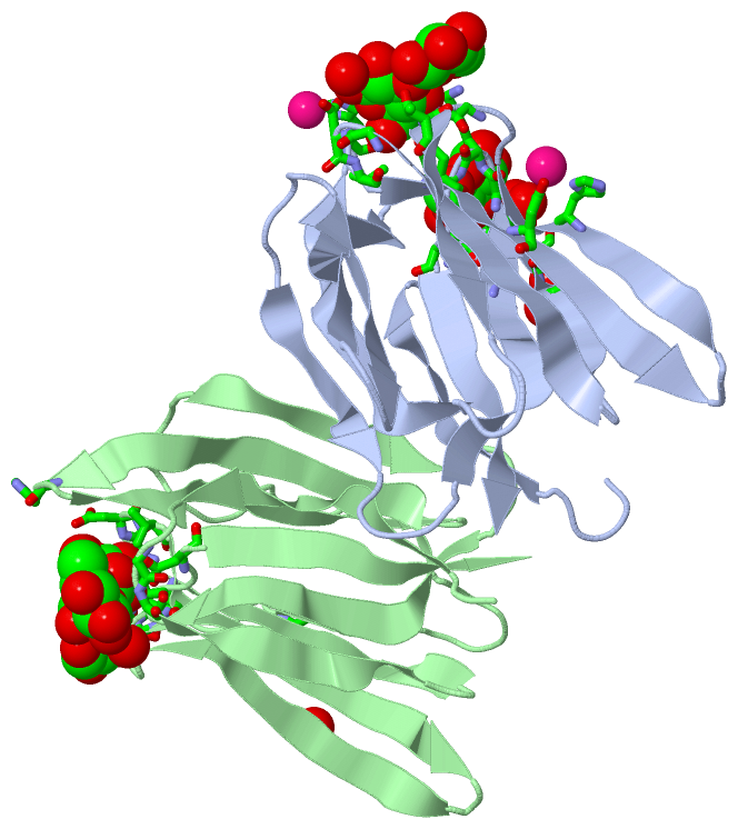
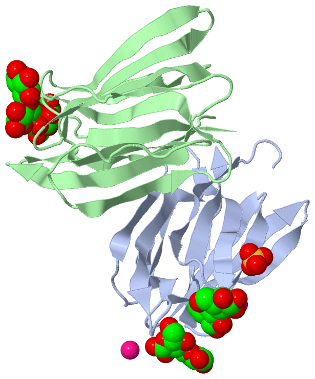
Still Images
|
||||||||||||||||
Databases
|
||||||||||||||||||||||||||||||||||||||||||||||||||||||||||||||||||||||||||||||||||||||||||||||||||||||||||||||||||||||||||||||||||||||||||||||||||||||||||||||||
Analysis Tools
|
|||||||||||||||||||||||||||||||||||||||||||||||||||||||||||||
Entries Sharing at Least One Protein Chain (UniProt ID)
Related Entries Specified in the PDB File
|
|