Show PDB file:   
         Plain Text   HTML   (compressed file size)
QuickSearch:   
by PDB,NDB,UniProt,PROSITE Code or Search Term(s)  
(-)Asym./Biol. Unit - manually
(-)Asym./Biol. Unit
(-)Asym./Biol. Unit - sites
collapse expand < >
Image Asym./Biol. Unit - manually
Asym./Biol. Unit - manually  (Jmol Viewer)
Image Asym./Biol. Unit
Asym./Biol. Unit  (Jmol Viewer)
Image Asym./Biol. Unit - sites
Asym./Biol. Unit - sites  (Jmol Viewer)

(-) Description

Title :  CRYSTAL STRUCTURE OF G ALPHA 13 IN COMPLEX WITH GDP
 
Authors :  M. R. Nance, J. J. G. Tesmer
Date :  11 Apr 05  (Deposition) - 15 Nov 05  (Release) - 13 Jul 11  (Revision)
Method :  X-RAY DIFFRACTION
Resolution :  2.00
Chains :  Asym./Biol. Unit :  A
Keywords :  Gtp-Binding, Lipoprotein, Membrane, Transducer, Signaling Protein (Keyword Search: [Gene Ontology, PubMed, Web (Google))
 
Reference :  B. Kreutz, D. M. Yau, M. R. Nance, S. Tanabe, J. J. Tesmer, T. Kozasa
A New Approach To Producing Functional G Alpha Subunits Yields The Activated And Deactivated Structures Of G Alpha(12/13) Proteins.
Biochemistry V. 45 167 2006
PubMed-ID: 16388592  |  Reference-DOI: 10.1021/BI051729T

(-) Compounds

Molecule 1 - G ALPHA I/13
    ChainsA
    EngineeredYES
    Expression SystemSPODOPTERA FRUGIPERDA
    Expression System CommonFALL ARMYWORM
    Expression System PlasmidPFASTBACHTA
    Expression System StrainSF9
    Expression System Taxid7108
    Expression System Vector TypeVIRUS
    FragmentN-TERMINAL RESIDUES 1-28 OF G ALPHA I FOLLOWED BY RESIDUES 47-377 OF G ALPHA 13
    Organism CommonHOUSE MOUSE
    Organism ScientificMUS MUSCULUS
    Organism Taxid10090

 Structural Features

(-) Chains, Units

  1
Asymmetric/Biological Unit A

Summary Information (see also Sequences/Alignments below)

(-) Ligands, Modified Residues, Ions  (1, 1)

Asymmetric/Biological Unit (1, 1)
No.NameCountTypeFull Name
1GDP1Ligand/IonGUANOSINE-5'-DIPHOSPHATE

(-) Sites  (1, 1)

Asymmetric Unit (1, 1)
No.NameEvidenceResiduesDescription
1AC1SOFTWAREGLU A:58 , SER A:59 , GLY A:60 , LYS A:61 , SER A:62 , THR A:63 , GLU A:172 , LEU A:197 , LEU A:198 , ARG A:200 , ASN A:291 , LYS A:292 , ASP A:294 , LEU A:295 , THR A:348 , ALA A:349 , ILE A:350 , HOH A:386 , HOH A:392 , HOH A:399 , HOH A:417 , HOH A:424BINDING SITE FOR RESIDUE GDP A 378

(-) SS Bonds  (0, 0)

(no "SS Bond" information available for 1ZCB)

(-) Cis Peptide Bonds  (0, 0)

(no "Cis Peptide Bond" information available for 1ZCB)

 Sequence-Structure Mapping

(-) SAPs(SNPs)/Variants  (0, 0)

(no "SAP(SNP)/Variant" information available for 1ZCB)

(-) PROSITE Motifs  (0, 0)

(no "PROSITE Motif" information available for 1ZCB)

(-) Exons   (0, 0)

(no "Exon" information available for 1ZCB)

(-) Sequences/Alignments

Asymmetric/Biological Unit
   Reformat: Number of residues per line =  ('0' or empty: single-line sequence representation)
  Number of residues per labelling interval =   
  UniProt sequence: complete  aligned part    
   Show mapping: SCOP domains CATH domains Pfam domains Secondary structure (by author)
SAPs(SNPs) PROSITE motifs Exons
(details for a mapped element are shown in a popup box when the mouse pointer rests over it)
Chain A from PDB  Type:PROTEIN  Length:318
 aligned with GNA13_MOUSE | P27601 from UniProtKB/Swiss-Prot  Length:377

    Alignment length:327
                                    55        65        75        85        95       105       115       125       135       145       155       165       175       185       195       205       215       225       235       245       255       265       275       285       295       305       315       325       335       345       355       365       
          GNA13_MOUSE    46 KRLVKILLLGAGESGKSTFLKQMRIIHGQDFDQRAREEFRPTIYSNVIKGMRVLVDAREKLHIPWGDNKNQLHGDKLMAFDTRAPMAAQGMVETRVFLQYLPAIRALWEDSGIQNAYDRRREFQLGESVKYFLDNLDKLGVPDYIPSQQDILLARRPTKGIHEYDFEIKNVPFKMVDVGGQRSERKRWFECFDSVTSILFLVSSSEFDQVLMEDRQTNRLTESLNIFETIVNNRVFSNVSIILFLNKTDLLEEKVQVVSIKDYFLEFEGDPHCLRDVQKFLVECFRGKRRDQQQRPLYHHFTTAINTENIRLVFRDVKDTILHDNLK 372
               SCOP domains -d1zcba2 A:47-75,A:202-372    d1zcba1 A:76-201 Transducin (alpha subunit), insertion domain                                                                 d1zcba2 A:47-75,A:202-37       2 Transducin (alpha subunit)                                                                                                                 SCOP domains
               CATH domains 1zcbA01 A:46-76,A:203-372      1zcbA02 A:77-202 GI Alpha 1, domain 2-like                                                                                    1zcbA01 A:46-76,A:203-3       72 P-loop containing nucleotide triphosphate hydrolases                                                                                      CATH domains
               Pfam domains -G-alpha-1zcbA01 A:47-372                                                                                                                                                                                                                                                                                                               Pfam domains
         Sec.struct. author ...eeeeee.....hhhhhhhhhhhhhh...hhhhhhhhhhhhhhhhhhhhhhhhhhhhhh.....hhhhhhhhhhhhh....hhhhhh...hhhhhhhhhhhhhhhhhhhhhhhhhhhhhhh....hhhhhhhhhhhhh......hhhhhhhh......eeeeeeee..eeeeeeee..-------.........eeeeeee......ee..eeeeehhhhhhhhhhhhhhhhhhh..eeeeeeehhhhhhhhh...hhhhh.........hhhhhhhhhhhhhhh......--..eeee....hhhhhhhhhhhhhhhhhhhhhh Sec.struct. author
                 SAPs(SNPs) --------------------------------------------------------------------------------------------------------------------------------------------------------------------------------------------------------------------------------------------------------------------------------------------------------------------------------------- SAPs(SNPs)
                    PROSITE --------------------------------------------------------------------------------------------------------------------------------------------------------------------------------------------------------------------------------------------------------------------------------------------------------------------------------------- PROSITE
                 Transcript --------------------------------------------------------------------------------------------------------------------------------------------------------------------------------------------------------------------------------------------------------------------------------------------------------------------------------------- Transcript
                 1zcb A  46 ARLVKILLLGAGESGKSTFLKQMRIIHGQDFDQRAREEFRPTIYSNVIKGMRVLVDAREKLHIPWGDNKNQLHGDKLMAFDTRAPMAAQGMVETRVFLQYLPAIRALWEDSGIQNAYDRRREFQLGESVKYFLDNLDKLGVPDYIPSQQDILLARRPTKGIHEYDFEIKNVPFKMVDVGG-------WFECFDSVTSILFLVSSSEFDQVLMEDRQTNRLTESLNIFETIVNNRVFSNVSIILFLNKTDLLEEKVQVVSIKDYFLEFEGDPHCLRDVQKFLVECFRGKRRDQQ--PLYHHFTTAINTENIRLVFRDVKDTILHDNLK 372
                                    55        65        75        85        95       105       115       125       135       145       155       165       175       185       195       205       215       225       235       245       255       265       275       285       295       305       315       325       335  |  | 345       355       365       
                                                                                                                                                                                                             225     233                                                                                                      338  |                               
                                                                                                                                                                                                                                                                                                                                 341                               

   Legend:   → Mismatch (orange background)
  - → Gap (green background, '-', border residues have a numbering label)
    → Modified Residue (blue background, lower-case, 'x' indicates undefined single-letter code, labelled with number + name)
  x → Chemical Group (purple background, 'x', labelled with number + name, e.g. ACE or NH2)
  extra numbering lines below/above indicate numbering irregularities and modified residue names etc., number ends below/above '|'

 Classification and Annotation

(-) SCOP Domains  (2, 2)

Asymmetric/Biological Unit

(-) CATH Domains  (2, 2)

Asymmetric/Biological Unit
(-)
Class: Alpha Beta (26913)

(-) Pfam Domains  (1, 1)

Asymmetric/Biological Unit

(-) Gene Ontology  (36, 36)

Asymmetric/Biological Unit(hide GO term definitions)
Chain A   (GNA13_MOUSE | P27601)
molecular function
    GO:0031752    D5 dopamine receptor binding    Interacting selectively and non-covalently with a D5 dopamine receptor.
    GO:0031683    G-protein beta/gamma-subunit complex binding    Interacting selectively and non-covalently with a complex of G-protein beta/gamma subunits.
    GO:0001664    G-protein coupled receptor binding    Interacting selectively and non-covalently with a G-protein coupled receptor.
    GO:0005525    GTP binding    Interacting selectively and non-covalently with GTP, guanosine triphosphate.
    GO:0003924    GTPase activity    Catalysis of the reaction: GTP + H2O = GDP + phosphate.
    GO:0019001    guanyl nucleotide binding    Interacting selectively and non-covalently with guanyl nucleotides, any compound consisting of guanosine esterified with (ortho)phosphate.
    GO:0046872    metal ion binding    Interacting selectively and non-covalently with any metal ion.
    GO:0000166    nucleotide binding    Interacting selectively and non-covalently with a nucleotide, any compound consisting of a nucleoside that is esterified with (ortho)phosphate or an oligophosphate at any hydroxyl group on the ribose or deoxyribose.
    GO:0005515    protein binding    Interacting selectively and non-covalently with any protein or protein complex (a complex of two or more proteins that may include other nonprotein molecules).
    GO:0004871    signal transducer activity    Conveys a signal across a cell to trigger a change in cell function or state. A signal is a physical entity or change in state that is used to transfer information in order to trigger a response.
    GO:0031702    type 1 angiotensin receptor binding    Interacting selectively and non-covalently with a type 1 angiotensin receptor.
biological process
    GO:0007186    G-protein coupled receptor signaling pathway    A series of molecular signals that proceeds with an activated receptor promoting the exchange of GDP for GTP on the alpha-subunit of an associated heterotrimeric G-protein complex. The GTP-bound activated alpha-G-protein then dissociates from the beta- and gamma-subunits to further transmit the signal within the cell. The pathway begins with receptor-ligand interaction, or for basal GPCR signaling the pathway begins with the receptor activating its G protein in the absence of an agonist, and ends with regulation of a downstream cellular process, e.g. transcription. The pathway can start from the plasma membrane, Golgi or nuclear membrane (PMID:24568158 and PMID:16902576).
    GO:0007266    Rho protein signal transduction    A series of molecular signals within the cell that are mediated by a member of the Rho family of proteins switching to a GTP-bound active state.
    GO:0031584    activation of phospholipase D activity    Any process that initiates the activity of inactive phospholipase D.
    GO:0007189    adenylate cyclase-activating G-protein coupled receptor signaling pathway    The series of molecular signals generated as a consequence of a G-protein coupled receptor binding to its physiological ligand, where the pathway proceeds through activation of adenylyl cyclase activity and a subsequent increase in the concentration of cyclic AMP (cAMP).
    GO:0001525    angiogenesis    Blood vessel formation when new vessels emerge from the proliferation of pre-existing blood vessels.
    GO:0001569    branching involved in blood vessel morphogenesis    The process of coordinated growth and sprouting of blood vessels giving rise to the organized vascular system.
    GO:0030154    cell differentiation    The process in which relatively unspecialized cells, e.g. embryonic or regenerative cells, acquire specialized structural and/or functional features that characterize the cells, tissues, or organs of the mature organism or some other relatively stable phase of the organism's life history. Differentiation includes the processes involved in commitment of a cell to a specific fate and its subsequent development to the mature state.
    GO:0001701    in utero embryonic development    The process whose specific outcome is the progression of the embryo in the uterus over time, from formation of the zygote in the oviduct, to birth. An example of this process is found in Mus musculus.
    GO:0035556    intracellular signal transduction    The process in which a signal is passed on to downstream components within the cell, which become activated themselves to further propagate the signal and finally trigger a change in the function or state of the cell.
    GO:0030168    platelet activation    A series of progressive, overlapping events triggered by exposure of the platelets to subendothelial tissue. These events include shape change, adhesiveness, aggregation, and release reactions. When carried through to completion, these events lead to the formation of a stable hemostatic plug.
    GO:0007204    positive regulation of cytosolic calcium ion concentration    Any process that increases the concentration of calcium ions in the cytosol.
    GO:0030334    regulation of cell migration    Any process that modulates the frequency, rate or extent of cell migration.
    GO:0008360    regulation of cell shape    Any process that modulates the surface configuration of a cell.
    GO:0007165    signal transduction    The cellular process in which a signal is conveyed to trigger a change in the activity or state of a cell. Signal transduction begins with reception of a signal (e.g. a ligand binding to a receptor or receptor activation by a stimulus such as light), or for signal transduction in the absence of ligand, signal-withdrawal or the activity of a constitutively active receptor. Signal transduction ends with regulation of a downstream cellular process, e.g. regulation of transcription or regulation of a metabolic process. Signal transduction covers signaling from receptors located on the surface of the cell and signaling via molecules located within the cell. For signaling between cells, signal transduction is restricted to events at and within the receiving cell.
    GO:0007264    small GTPase mediated signal transduction    Any series of molecular signals in which a small monomeric GTPase relays one or more of the signals.
cellular component
    GO:0031526    brush border membrane    The portion of the plasma membrane surrounding the brush border.
    GO:0005737    cytoplasm    All of the contents of a cell excluding the plasma membrane and nucleus, but including other subcellular structures.
    GO:0005829    cytosol    The part of the cytoplasm that does not contain organelles but which does contain other particulate matter, such as protein complexes.
    GO:0070062    extracellular exosome    A vesicle that is released into the extracellular region by fusion of the limiting endosomal membrane of a multivesicular body with the plasma membrane. Extracellular exosomes, also simply called exosomes, have a diameter of about 40-100 nm.
    GO:0005925    focal adhesion    Small region on the surface of a cell that anchors the cell to the extracellular matrix and that forms a point of termination of actin filaments.
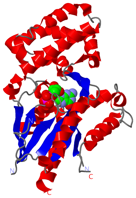
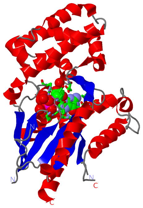
    GO:0005834    heterotrimeric G-protein complex    Any of a family of heterotrimeric GTP-binding and hydrolyzing proteins; they belong to a superfamily of GTPases that includes monomeric proteins such as EF-Tu and RAS. Heterotrimeric G-proteins consist of three subunits; the alpha subunit contains the guanine nucleotide binding site and possesses GTPase activity; the beta and gamma subunits are tightly associated and function as a beta-gamma heterodimer; extrinsic plasma membrane proteins (cytoplasmic face) that function as a complex to transduce signals from G-protein coupled receptors to an effector protein.
    GO:0042470    melanosome    A tissue-specific, membrane-bounded cytoplasmic organelle within which melanin pigments are synthesized and stored. Melanosomes are synthesized in melanocyte cells.
    GO:0016020    membrane    A lipid bilayer along with all the proteins and protein complexes embedded in it an attached to it.
    GO:0005634    nucleus    A membrane-bounded organelle of eukaryotic cells in which chromosomes are housed and replicated. In most cells, the nucleus contains all of the cell's chromosomes except the organellar chromosomes, and is the site of RNA synthesis and processing. In some species, or in specialized cell types, RNA metabolism or DNA replication may be absent.
    GO:0005886    plasma membrane    The membrane surrounding a cell that separates the cell from its external environment. It consists of a phospholipid bilayer and associated proteins.

 Visualization

(-) Interactive Views

Asymmetric/Biological Unit
  Complete Structure
    Jena3D(integrated viewing of ligand, site, SAP, PROSITE, SCOP information)
    WebMol | AstexViewer[tm]@PDBe
(Java Applets, require no local installation except for Java; loading may be slow)
    STRAP
(Java WebStart application, automatic local installation, requires Java; full application with system access!)
    RasMol
(require local installation)
    Molscript (VRML)
(requires installation of a VRML viewer; select preferred view via VRML and generate a mono or stereo PDF format file)
 
  Ligands, Modified Residues, Ions
    GDP  [ RasMol | Jena3D ]  +environment [ RasMol | Jena3D ]
 
  Sites
    AC1  [ RasMol ]  +environment [ RasMol ]
 
  Cis Peptide Bonds
(no "Cis Peptide Bonds" information available for 1zcb)
 

(-) Still Images

Jmol
  protein: cartoon or spacefill or dots and stick; nucleic acid: cartoon and stick; ligands: spacefill; active site: stick
Molscript
  protein, nucleic acid: cartoon; ligands: spacefill; active site: ball and stick

 Databases and Analysis Tools

(-) Databases

Access by PDB/NDB ID
  1zcb
    Family and Domain InformationProDom | SYSTERS
    General Structural InformationGlycoscienceDB | MMDB | NDB | OCA | PDB | PDBe | PDBj | PDBsum | PDBWiki | PQS | PROTEOPEDIA
    Orientation in MembranesOPM
    Protein SurfaceSURFACE
    Secondary StructureDSSP (structure derived) | HSSP (homology derived)
    Structural GenomicsGeneCensus
    Structural NeighboursCE | VAST
    Structure ClassificationCATH | Dali | SCOP
    Validation and Original DataBMRB Data View | BMRB Restraints Grid | EDS | PROCHECK | RECOORD | WHAT_CHECK
 
Access by UniProt ID/Accession number
  GNA13_MOUSE | P27601
    Comparative Protein Structure ModelsModBase
    Genomic InformationEnsembl
    Protein-protein InteractionDIP
    Sequence, Family and Domain InformationInterPro | Pfam | SMART | UniProtKB/SwissProt
 
Access by Enzyme Classificator   (EC Number)
  (no 'Enzyme Classificator' available)
    General Enzyme InformationBRENDA | EC-PDB | Enzyme | IntEnz
    PathwayKEGG | MetaCyc
 
Access by Disease Identifier   (MIM ID)
  (no 'MIM ID' available)
    Disease InformationOMIM
 
Access by GenAge ID
  (no 'GenAge ID' available)
    Age Related InformationGenAge

(-) Analysis Tools

Access by PDB/NDB ID
    Domain InformationXDom
    Interatomic Contacts of Structural UnitsCSU
    Ligand-protein ContactsLPC
    Protein CavitiescastP
    Sequence and Secondary StructurePDBCartoon
    Structure AlignmentSTRAP(Java WebStart application, automatic local installation, requires Java; full application with system access!)
    Structure and Sequence BrowserSTING
 
Access by UniProt ID/Accession number
  GNA13_MOUSE | P27601
    Protein Disorder PredictionDisEMBL | FoldIndex | GLOBPLOT (for more information see DisProt)

 Related Entries

(-) Entries Sharing at Least One Protein Chain (UniProt ID)

UniProtKB/Swiss-Prot
        GNA13_MOUSE | P276011shz 3ab3 3cx6 3cx7 3cx8

(-) Related Entries Specified in the PDB File

1zca CRYSTAL STRUCTURE OF G ALPHA 12 IN COMPLEX WITH GDP, MG2+ AND ALF4-