|
|
|
|
 Description
|
|
Compounds
|
||||||||||||||||||||||||||||||||||||
Chains, Units
Summary Information (see also Sequences/Alignments below) |
Ligands, Modified Residues, Ions (2, 14)
Asymmetric Unit (2, 14)
|
Sites (10, 10)
Asymmetric Unit (10, 10)
|
SS Bonds (0, 0)| (no "SS Bond" information available for 1XIO) |
Cis Peptide Bonds (0, 0)| (no "Cis Peptide Bond" information available for 1XIO) |
SAPs(SNPs)/Variants (0, 0)| (no "SAP(SNP)/Variant" information available for 1XIO) |
PROSITE Motifs (0, 0)| (no "PROSITE Motif" information available for 1XIO) |
Exons (0, 0)| (no "Exon" information available for 1XIO) |
Sequences/Alignments
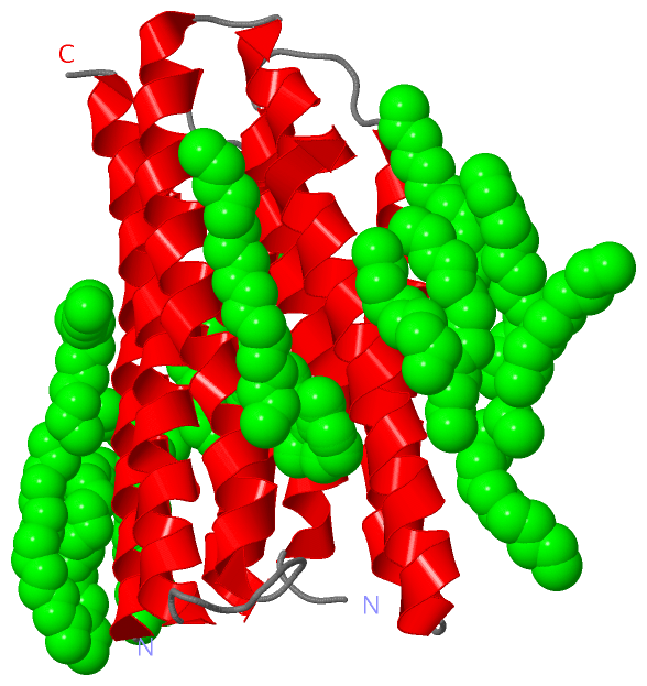
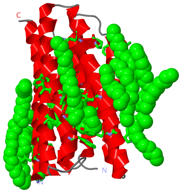
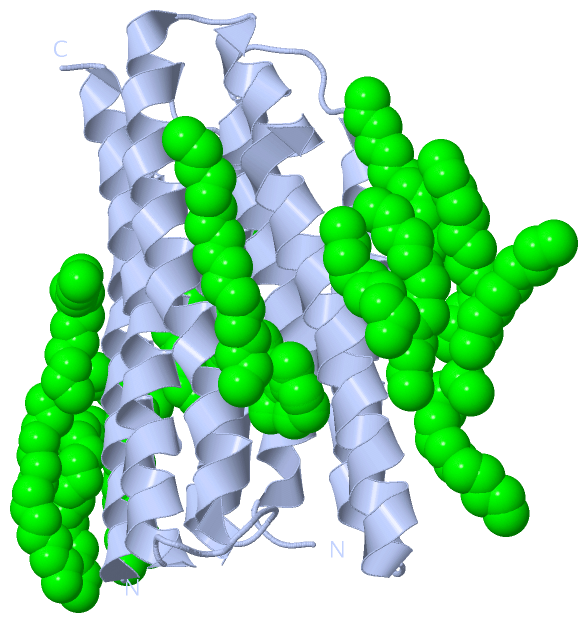
Asymmetric UnitChain A from PDB Type:PROTEIN Length:217 aligned with Q8YSC4_NOSS1 | Q8YSC4 from UniProtKB/TrEMBL Length:261 Alignment length:226 10 20 30 40 50 60 70 80 90 100 110 120 130 140 150 160 170 180 190 200 210 220 Q8YSC4_NOSS1 1 MNLESLLHWIYVAGMTIGALHFWSLSRNPRGVPQYEYLVAMFIPIWSGLAYMAMAIDQGKVEAAGQIAHYARYIDWMVTTPLLLLSLSWTAMQFIKKDWTLIGFLMSTQIVVITSGLIADLSERDWVRYLWYICGVCAFLIILWGIWNPLRAKTRTQSSELANLYDKLVTYFTVLWIGYPIVWIIGPSGFGWINQTIDTFLFCLLPFFSKVGFSFLDLHGLRNLND 226 SCOP domains d1xioa_ A: Sensory rhodopsin SCOP domains CATH domains 1xioA00 A:1-226 Rhopdopsin 7-helix transmembrane proteins CATH domains Pfam domains ----Bac_rhodopsin-1xioA01 A:5-226 Pfam domains SAPs(SNPs) ---------------------------------------------------------------------------------------------------------------------------------------------------------------------------------------------------------------------------------- SAPs(SNPs) PROSITE ---------------------------------------------------------------------------------------------------------------------------------------------------------------------------------------------------------------------------------- PROSITE Transcript ---------------------------------------------------------------------------------------------------------------------------------------------------------------------------------------------------------------------------------- Transcript 1xio A 1 MNLESLLHWIYVAGMTIGALHFWSLSRNPRGVPQYEYLVAMFIPIWSGLAYMAMAID---------IAHYARYIDWMVTTPLLLLSLSWTAMQFIKKDWTLIGFLMSTQIVVITSGLIADLSERDWVRYLWYICGVCAFLIILWGIWNPLRAKTRTQSSELANLYDKLVTYFTVLWIGYPIVWIIGPSGFGWINQTIDTFLFCLLPFFSKVGFSFLDLHGLRNLND 226 10 20 30 40 50 | - | 70 80 90 100 110 120 130 140 150 160 170 180 190 200 210 220 57 67
|
||||||||||||||||||||
SCOP Domains (1, 1)
Asymmetric Unit
|
CATH Domains (1, 1)
Asymmetric Unit
 |
Pfam Domains (1, 1)
Asymmetric Unit
|
Gene Ontology (5, 5)|
Asymmetric Unit(hide GO term definitions) Chain A (Q8YSC4_NOSS1 | Q8YSC4)
|
||||||||||||||||||||||||||||||||||||||||||||||||
Interactive Views
|
|||||||||||||||||||||||||||||||||||||||||||||||||||||||||||||||||||||||||||||||||||||||||||||||||||||||||||||||||||||||||||||||||||||||||||||||||||||||||||||||||||||||||||||||||||||||||||||||||||||||||||||||||||
Still Images
|
||||||||||||||||
Databases
|
||||||||||||||||||||||||||||||||||||||||||||||||||||||||||||||||||||||||||||||||||||||||||||||||||||||||||||||||||||||||||||||||||||||||||||||||||||||||||||||||
Analysis Tools
|
|||||||||||||||||||||||||||||||||||||||||||||||||||||||||||||
Entries Sharing at Least One Protein Chain (UniProt ID)
Related Entries Specified in the PDB File
|
|