|
|
|
|
 Description
|
|
Compounds
|
||||||||||||||||||||||||||||||||||||||||||||||||
Chains, Units
Summary Information (see also Sequences/Alignments below) |
Ligands, Modified Residues, Ions (1, 4)
Asymmetric Unit (1, 4)
|
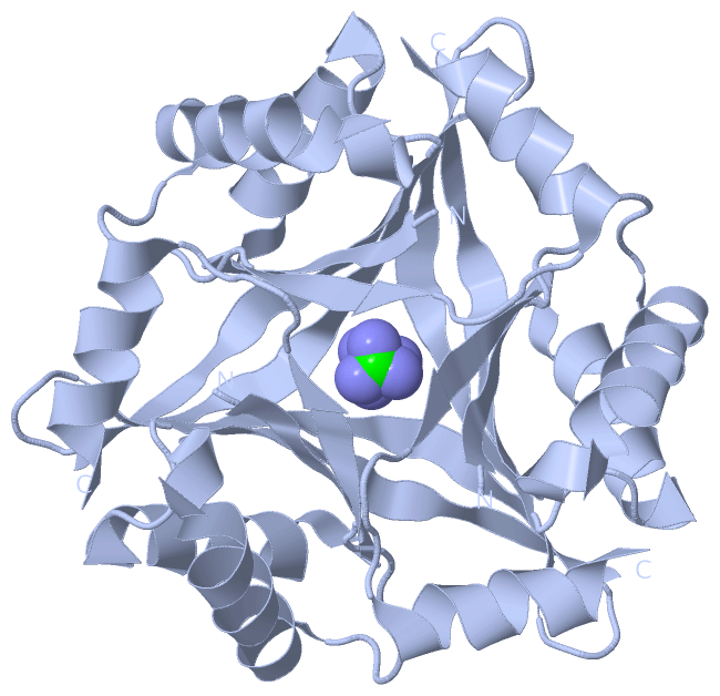
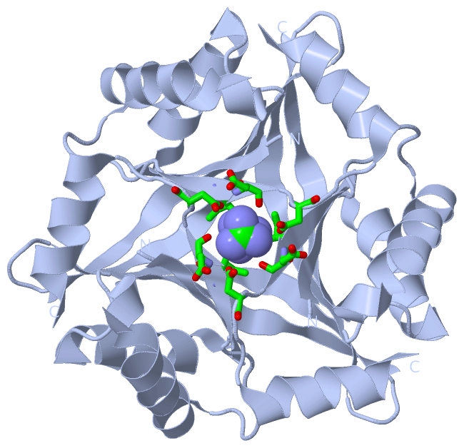
Sites (4, 4)
Asymmetric Unit (4, 4)
|
SS Bonds (0, 0)| (no "SS Bond" information available for 1UMJ) |
Cis Peptide Bonds (0, 0)| (no "Cis Peptide Bond" information available for 1UMJ) |
SAPs(SNPs)/Variants (0, 0)| (no "SAP(SNP)/Variant" information available for 1UMJ) |
PROSITE Motifs (0, 0)| (no "PROSITE Motif" information available for 1UMJ) |
Exons (0, 0)| (no "Exon" information available for 1UMJ) |
Sequences/Alignments
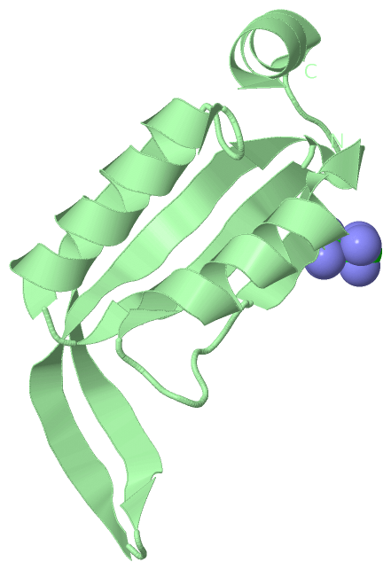
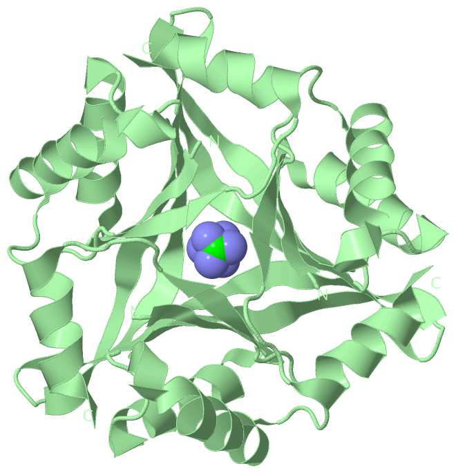
Asymmetric UnitChain A from PDB Type:PROTEIN Length:101 aligned with CUTA_PYRHO | O58720 from UniProtKB/Swiss-Prot Length:102 Alignment length:101 10 20 30 40 50 60 70 80 90 100 CUTA_PYRHO 1 MIIVYTTFPDWESAEKVVKTLLKERLIACANLREHRAFYWWEGKIEEDKEVGAILKTREDLWEELKERIKELHPYDVPAIIRIDVDDVNEDYLKWLIEETK 101 SCOP domains d1umja_ A: Cut A1 SCOP domains CATH domains 1umjA00 A:1-101 [code=3.30.70.830, no name defined] CATH domains Pfam domains ----------------------------------------------------------------------------------------------------- Pfam domains SAPs(SNPs) ----------------------------------------------------------------------------------------------------- SAPs(SNPs) PROSITE ----------------------------------------------------------------------------------------------------- PROSITE Transcript ----------------------------------------------------------------------------------------------------- Transcript 1umj A 1 MIIVYTTFPDWESAEKVVKTLLKERLIACANLREHRAFYWWEGKIEEDKEVGAILKTREDLWEELKERIKELHPYDVPAIIRIDVDDVNEDYLKWLIEETK 101 10 20 30 40 50 60 70 80 90 100 Chain B from PDB Type:PROTEIN Length:101 aligned with CUTA_PYRHO | O58720 from UniProtKB/Swiss-Prot Length:102 Alignment length:101 10 20 30 40 50 60 70 80 90 100 CUTA_PYRHO 1 MIIVYTTFPDWESAEKVVKTLLKERLIACANLREHRAFYWWEGKIEEDKEVGAILKTREDLWEELKERIKELHPYDVPAIIRIDVDDVNEDYLKWLIEETK 101 SCOP domains d1umjb_ B: Cut A1 SCOP domains CATH domains 1umjB00 B:1-101 [code=3.30.70.830, no name defined] CATH domains Pfam domains (1) CutA1-1umjB01 B:1-101 Pfam domains (1) Pfam domains (2) CutA1-1umjB02 B:1-101 Pfam domains (2) SAPs(SNPs) ----------------------------------------------------------------------------------------------------- SAPs(SNPs) PROSITE ----------------------------------------------------------------------------------------------------- PROSITE Transcript ----------------------------------------------------------------------------------------------------- Transcript 1umj B 1 MIIVYTTFPDWESAEKVVKTLLKERLIACANLREHRAFYWWEGKIEEDKEVGAILKTREDLWEELKERIKELHPYDVPAIIRIDVDDVNEDYLKWLIEETK 101 10 20 30 40 50 60 70 80 90 100
|
||||||||||||||||||||
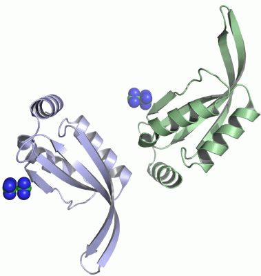
SCOP Domains (1, 2)
Asymmetric Unit
 |
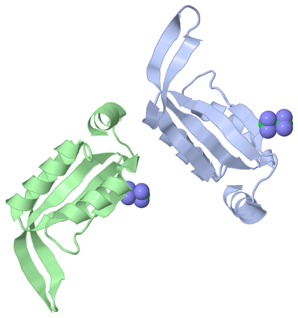
CATH Domains (1, 2)
Asymmetric Unit
|
Pfam Domains (1, 2)
Asymmetric Unit
|
Gene Ontology (3, 3)|
Asymmetric Unit(hide GO term definitions) Chain A,B (CUTA_PYRHO | O58720)
|
||||||||||||||||||||||||||||||||||||
Interactive Views
|
|||||||||||||||||||||||||||||||||||||||||||||||||||||||||||||||||||||||||||||||||||||||||||||||||||||||||||||||||||||||||||||||||||||||||||||||||||||||||||||||||||||||
Still Images
|
||||||||||||||||
Databases
|
||||||||||||||||||||||||||||||||||||||||||||||||||||||||||||||||||||||||||||||||||||||||||||||||||||||||||||||||||||||||||||||||||||||||||||||||||||||||||||||||
Analysis Tools
|
|||||||||||||||||||||||||||||||||||||||||||||||||||||||||||||
Entries Sharing at Least One Protein Chain (UniProt ID)
Related Entries Specified in the PDB File
|
|