|
|
|
|
 Description
|
|
Compounds
|
||||||||||||||||||||||||||||||||||||
Chains, Units
Summary Information (see also Sequences/Alignments below) |
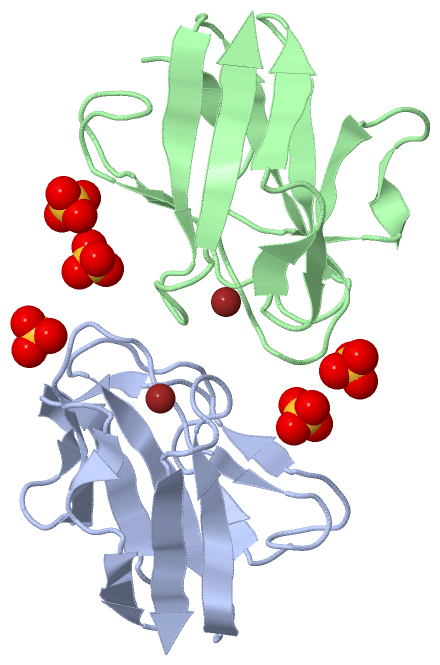
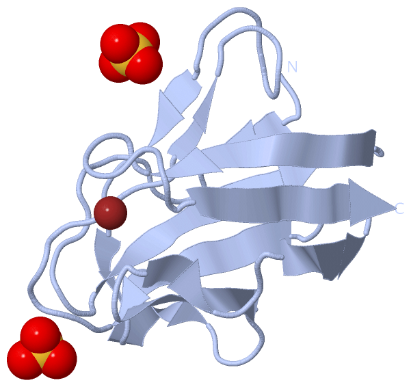
Ligands, Modified Residues, Ions (2, 7)| Asymmetric Unit (2, 7) Biological Unit 1 (1, 2) Biological Unit 2 (1, 3) |
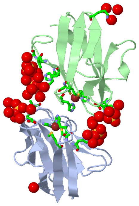
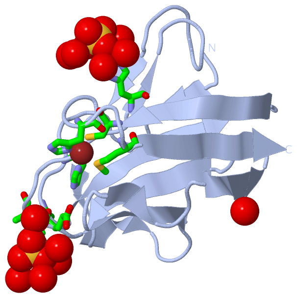
Sites (7, 7)
Asymmetric Unit (7, 7)
|
SS Bonds (0, 0)| (no "SS Bond" information available for 1SFD) |
Cis Peptide Bonds (0, 0)| (no "Cis Peptide Bond" information available for 1SFD) |
SAPs(SNPs)/Variants (0, 0)| (no "SAP(SNP)/Variant" information available for 1SFD) |
PROSITE Motifs (1, 2)
Asymmetric Unit (1, 2)
|
||||||||||||||||||||||||||||||||||||||||||||||||||||||||||||||||||||||||
Exons (0, 0)| (no "Exon" information available for 1SFD) |
Sequences/Alignments
Asymmetric UnitChain A from PDB Type:PROTEIN Length:105 aligned with AMCY_PARDE | P22364 from UniProtKB/Swiss-Prot Length:131 Alignment length:105 36 46 56 66 76 86 96 106 116 126 AMCY_PARDE 27 DKATIPSESPFAAAEVADGAIVVDIAKMKYETPELHVKVGDTVTWINREAMPHNVHFVAGVLGEAALKGPMMKKEQAYSLTFTEAGTYDYHCTPHPFMRGKVVVE 131 SCOP domains d1sfda_ A: Amicyanin SCOP domains CATH domains 1sfdA00 A:1-105 Cupredoxins - blue copper proteins CATH domains Pfam domains --------------------------------------------------------------------------------------------------------- Pfam domains SAPs(SNPs) --------------------------------------------------------------------------------------------------------- SAPs(SNPs) PROSITE ------------------------------------------------------------------------------------COPPER_BLUE ------- PROSITE Transcript --------------------------------------------------------------------------------------------------------- Transcript 1sfd A 1 DKATIPSESPFAAAEVADGAIVVDIAKMKYETPELHVKVGDTVTWINREAMPHNVHFVAGVLGEAALKGPMMKKEQAYSLTFTEAGTYDYHCTFHPFMRGKVVVE 105 10 20 30 40 50 60 70 80 90 100 Chain B from PDB Type:PROTEIN Length:105 aligned with AMCY_PARDE | P22364 from UniProtKB/Swiss-Prot Length:131 Alignment length:105 36 46 56 66 76 86 96 106 116 126 AMCY_PARDE 27 DKATIPSESPFAAAEVADGAIVVDIAKMKYETPELHVKVGDTVTWINREAMPHNVHFVAGVLGEAALKGPMMKKEQAYSLTFTEAGTYDYHCTPHPFMRGKVVVE 131 SCOP domains d1sfdb_ B: Amicyanin SCOP domains CATH domains 1sfdB00 B:1-105 Cupredoxins - blue copper proteins CATH domains Pfam domains (1) ----------------Copper-bind-1sfdB01 B:17-105 Pfam domains (1) Pfam domains (2) ----------------Copper-bind-1sfdB02 B:17-105 Pfam domains (2) SAPs(SNPs) --------------------------------------------------------------------------------------------------------- SAPs(SNPs) PROSITE ------------------------------------------------------------------------------------COPPER_BLUE ------- PROSITE Transcript --------------------------------------------------------------------------------------------------------- Transcript 1sfd B 1 DKATIPSESPFAAAEVADGAIVVDIAKMKYETPELHVKVGDTVTWINREAMPHNVHFVAGVLGEAALKGPMMKKEQAYSLTFTEAGTYDYHCTFHPFMRGKVVVE 105 10 20 30 40 50 60 70 80 90 100
|
||||||||||||||||||||
SCOP Domains (1, 2)
Asymmetric Unit
 |
CATH Domains (1, 2)
Asymmetric Unit
|
Pfam Domains (1, 2)
Asymmetric Unit
|
Gene Ontology (5, 5)|
Asymmetric Unit(hide GO term definitions) Chain A,B (AMCY_PARDE | P22364)
|
||||||||||||||||||||||||||||||||||||||||||||||||
Interactive Views
|
||||||||||||||||||||||||||||||||||||||||||||||||||||||||||||||||||||||||||||||||||||||||||||||||||||||||||||||||||||||||||||||||||||||||||||||||||||||||||||||||||||||||||||||||||||||||||||||
Still Images
|
||||||||||||||||
Databases
|
||||||||||||||||||||||||||||||||||||||||||||||||||||||||||||||||||||||||||||||||||||||||||||||||||||||||||||||||||||||||||||||||||||||||||||||||||||||||||||||||
Analysis Tools
|
|||||||||||||||||||||||||||||||||||||||||||||||||||||||||||||
Entries Sharing at Least One Protein Chain (UniProt ID)
Related Entries Specified in the PDB File
|
|