Show PDB file:   
         Plain Text   HTML   (compressed file size)
QuickSearch:   
by PDB,NDB,UniProt,PROSITE Code or Search Term(s)  
(-)Biol.Unit 1 - manually
(-)Asym.Unit - manually
(-)Asymmetric Unit
(-)Asym. Unit - sites
(-)Biological Unit 1
(-)Biol. Unit 1 - sites
(-)Biological Unit 2
collapse expand < >
Image Biol.Unit 1 - manually
Biol.Unit 1 - manually  (Jmol Viewer)
Image Asym.Unit - manually
Asym.Unit - manually  (Jmol Viewer)
Image Asymmetric Unit
Asymmetric Unit  (Jmol Viewer)
Image Asym. Unit - sites
Asym. Unit - sites  (Jmol Viewer)
Image Biological Unit 1
Biological Unit 1  (Jmol Viewer)
Image Biol. Unit 1 - sites
Biol. Unit 1 - sites  (Jmol Viewer)
Image Biological Unit 2
Biological Unit 2  (Jmol Viewer)

(-) Description

Title :  RUBRERYTHRIN
 
Authors :  F. Demare, D. M. Kurtz, P. Nordlund
Date :  26 Apr 96  (Deposition) - 15 May 97  (Release) - 13 Jul 11  (Revision)
Method :  X-RAY DIFFRACTION
Resolution :  2.10
Chains :  Asym. Unit :  A
Biol. Unit 1:  A  (2x)
Biol. Unit 2:  A  (4x)
Keywords :  Electron Transport, Iron, Ferroxidase (Keyword Search: [Gene Ontology, PubMed, Web (Google))
 
Reference :  F. Demare, D. M. Kurtz Jr. , P. Nordlund
The Structure Of Desulfovibrio Vulgaris Rubrerythrin Reveal A Unique Combination Of Rubredoxin-Like Fes4 And Ferritin-Like Diiron Domains.
Nat. Struct. Biol. V. 3 539 1996
PubMed-ID: 8646540  |  Reference-DOI: 10.1038/NSB0696-539

(-) Compounds

Molecule 1 - RUBRERYTHRIN
    ChainsA
    Organism ScientificDESULFOVIBRIO VULGARIS SUBSP. VULGARIS STR. HILDENBOROUGH
    Organism Taxid882
    StrainHILDENBOROUGH

 Structural Features

(-) Chains, Units

  1
Asymmetric Unit A
Biological Unit 1 (2x)A
Biological Unit 2 (4x)A

Summary Information (see also Sequences/Alignments below)

(-) Ligands, Modified Residues, Ions  (1, 3)

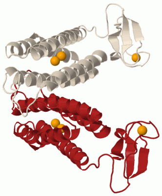
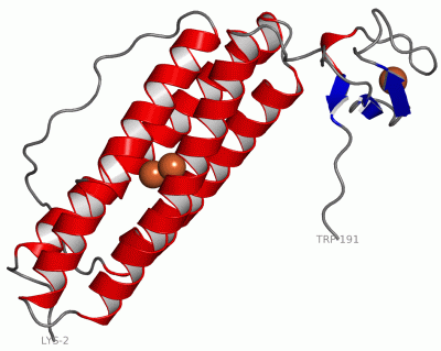
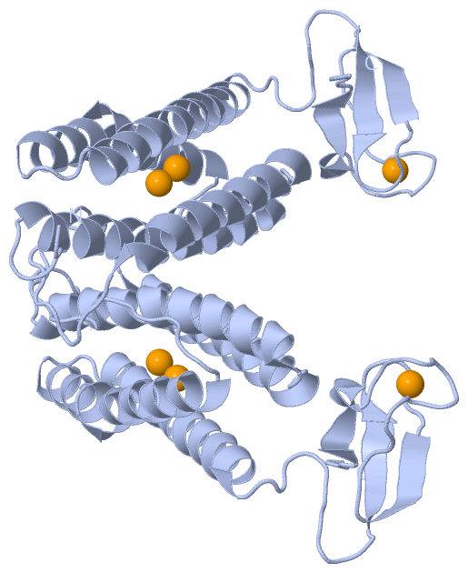
Asymmetric Unit (1, 3)
No.NameCountTypeFull Name
1FE3Ligand/IonFE (III) ION
Biological Unit 1 (0, 0)
No.NameCountTypeFull Name
1FE-1Ligand/IonFE (III) ION
Biological Unit 2 (0, 0)
No.NameCountTypeFull Name
1FE-1Ligand/IonFE (III) ION

(-) Sites  (3, 3)

Asymmetric Unit (3, 3)
No.NameEvidenceResiduesDescription
1AC1SOFTWAREGLU A:53 , GLU A:94 , GLU A:128 , HIS A:131 , FE A:601 , HOH A:680BINDING SITE FOR RESIDUE FE A 600
2AC2SOFTWAREGLU A:20 , GLU A:53 , GLU A:97 , GLU A:128 , FE A:600 , HOH A:680BINDING SITE FOR RESIDUE FE A 601
3AC3SOFTWARECYS A:158 , CYS A:161 , CYS A:174 , CYS A:177BINDING SITE FOR RESIDUE FE A 401

(-) SS Bonds  (0, 0)

(no "SS Bond" information available for 1RYT)

(-) Cis Peptide Bonds  (0, 0)

(no "Cis Peptide Bond" information available for 1RYT)

 Sequence-Structure Mapping

(-) SAPs(SNPs)/Variants  (0, 0)

(no "SAP(SNP)/Variant" information available for 1RYT)

(-) PROSITE Motifs  (2, 2)

Asymmetric Unit (2, 2)
 PROSITEUniProtKBPDB
No.IDACDescriptionIDLocationCountLocation
1FERRITIN_LIKEPS50905 Ferritin-like diiron domain profile.RUBY_DESVH3-146  1A:3-146
2RUBREDOXIN_LIKEPS50903 Rubredoxin-like domain profile.RUBY_DESVH153-187  1A:153-187
Biological Unit 1 (2, 4)
 PROSITEUniProtKBPDB
No.IDACDescriptionIDLocationCountLocation
1FERRITIN_LIKEPS50905 Ferritin-like diiron domain profile.RUBY_DESVH3-146  2A:3-146
2RUBREDOXIN_LIKEPS50903 Rubredoxin-like domain profile.RUBY_DESVH153-187  2A:153-187
Biological Unit 2 (2, 8)
 PROSITEUniProtKBPDB
No.IDACDescriptionIDLocationCountLocation
1FERRITIN_LIKEPS50905 Ferritin-like diiron domain profile.RUBY_DESVH3-146  4A:3-146
2RUBREDOXIN_LIKEPS50903 Rubredoxin-like domain profile.RUBY_DESVH153-187  4A:153-187

(-) Exons   (0, 0)

(no "Exon" information available for 1RYT)

(-) Sequences/Alignments

Asymmetric Unit
   Reformat: Number of residues per line =  ('0' or empty: single-line sequence representation)
  Number of residues per labelling interval =   
  UniProt sequence: complete  aligned part    
   Show mapping: SCOP domains CATH domains Pfam domains Secondary structure (by author)
SAPs(SNPs) PROSITE motifs Exons
(details for a mapped element are shown in a popup box when the mouse pointer rests over it)
Chain A from PDB  Type:PROTEIN  Length:190
 aligned with RUBY_DESVH | P24931 from UniProtKB/Swiss-Prot  Length:191

    Alignment length:190
                                    11        21        31        41        51        61        71        81        91       101       111       121       131       141       151       161       171       181       191
           RUBY_DESVH     2 KSLKGSRTEKNILTAFAGESQARNRYNYFGGQAKKDGFVQISDIFAETADQEREHAKRLFKFLEGGDLEIVAAFPAGIIADTHANLIASAAGEHHEYTEMYPSFARIAREEGYEEIARVFASIAVAEEFHEKRFLDFARNIKEGRVFLREQATKWRCRNCGYVHEGTGAPELCPACAHPKAHFELLGINW 191
               SCOP domains d1ryta1 A:2-147 Rubrerythrin, N-terminal domain                                                                                                   d1ryta2 A:148-191                            SCOP domains
               CATH domains 1rytA01 A:2-146  [code=1.20.1260.10, no name defined]                                                                                            1rytA02 A:147-191                             CATH domains
               Pfam domains ---------Rubrerythrin-1rytA01 A:11-139                                                                                                    ---------------------------------------------------- Pfam domains
         Sec.struct. author ......hhhhhhhhhhhhhhhhhhhhhhhhhhhhhh..hhhhhhhhhhhhhhhhhhhhhhhh...................hhhhhhhhhhhhhhhhh.hhhhhhhhhhh...hhhhhhhhhhhhhhhhhhhhhhhhhhhhh..........eeeee.....eeee.............hhheee..... Sec.struct. author
                 SAPs(SNPs) ---------------------------------------------------------------------------------------------------------------------------------------------------------------------------------------------- SAPs(SNPs)
                    PROSITE -FERRITIN_LIKE  PDB: A:3-146 UniProt: 3-146                                                                                                      ------RUBREDOXIN_LIKE  PDB: A:153-187    ---- PROSITE
                 Transcript ---------------------------------------------------------------------------------------------------------------------------------------------------------------------------------------------- Transcript
                 1ryt A   2 KSLKGSRTEKNILTAFAGESQARNRYNYFGGQAKKDGFVQISDIFAETADQEREHAKRLFKFLEGGDLEIVAAFPAGIIADTHANLIASAAGEHHEYTEMYPSFARIAREEGYEEIARVFASIAVAEEFHEKRFLDFARNIKEGRVFLREQATKWRCRNCGYVHEGTGAPELCPACAHPKAHFELLGINW 191
                                    11        21        31        41        51        61        71        81        91       101       111       121       131       141       151       161       171       181       191

   Legend:   → Mismatch (orange background)
  - → Gap (green background, '-', border residues have a numbering label)
    → Modified Residue (blue background, lower-case, 'x' indicates undefined single-letter code, labelled with number + name)
  x → Chemical Group (purple background, 'x', labelled with number + name, e.g. ACE or NH2)
  extra numbering lines below/above indicate numbering irregularities and modified residue names etc., number ends below/above '|'

 Classification and Annotation

(-) SCOP Domains  (2, 2)

Asymmetric Unit

(-) CATH Domains  (2, 2)

Asymmetric Unit
(-)
Class: Mainly Beta (13760)

(-) Pfam Domains  (1, 1)

Asymmetric Unit
(-)
Clan: Ferritin (185)

(-) Gene Ontology  (6, 6)

Asymmetric Unit(hide GO term definitions)
Chain A   (RUBY_DESVH | P24931)
molecular function
    GO:0005506    iron ion binding    Interacting selectively and non-covalently with iron (Fe) ions.
    GO:0046872    metal ion binding    Interacting selectively and non-covalently with any metal ion.
    GO:0016491    oxidoreductase activity    Catalysis of an oxidation-reduction (redox) reaction, a reversible chemical reaction in which the oxidation state of an atom or atoms within a molecule is altered. One substrate acts as a hydrogen or electron donor and becomes oxidized, while the other acts as hydrogen or electron acceptor and becomes reduced.
biological process
    GO:0055114    oxidation-reduction process    A metabolic process that results in the removal or addition of one or more electrons to or from a substance, with or without the concomitant removal or addition of a proton or protons.
cellular component
    GO:0005575    cellular_component    The part of a cell, extracellular environment or virus in which a gene product is located. A gene product may be located in one or more parts of a cell and its location may be as specific as a particular macromolecular complex, that is, a stable, persistent association of macromolecules that function together.
    GO:0005737    cytoplasm    All of the contents of a cell excluding the plasma membrane and nucleus, but including other subcellular structures.

 Visualization

(-) Interactive Views

Asymmetric Unit
  Complete Structure
    Jena3D(integrated viewing of ligand, site, SAP, PROSITE, SCOP information)
    WebMol | AstexViewer[tm]@PDBe
(Java Applets, require no local installation except for Java; loading may be slow)
    STRAP
(Java WebStart application, automatic local installation, requires Java; full application with system access!)
    RasMol
(require local installation)
    Molscript (VRML)
(requires installation of a VRML viewer; select preferred view via VRML and generate a mono or stereo PDF format file)
 
  Ligands, Modified Residues, Ions
    FE  [ RasMol | Jena3D ]  +environment [ RasMol | Jena3D ]
 
  Sites
    AC1  [ RasMol ]  +environment [ RasMol ]
    AC2  [ RasMol ]  +environment [ RasMol ]
    AC3  [ RasMol ]  +environment [ RasMol ]
 
  Cis Peptide Bonds
(no "Cis Peptide Bonds" information available for 1ryt)
 
Biological Units
  Complete Structure
    Biological Unit 1  [ Jena3D ]
    Biological Unit 2  [ Jena3D ]

(-) Still Images

Jmol
  protein: cartoon or spacefill or dots and stick; nucleic acid: cartoon and stick; ligands: spacefill; active site: stick
Molscript
  protein, nucleic acid: cartoon; ligands: spacefill; active site: ball and stick

 Databases and Analysis Tools

(-) Databases

Access by PDB/NDB ID
  1ryt
    Family and Domain InformationProDom | SYSTERS
    General Structural InformationGlycoscienceDB | MMDB | NDB | OCA | PDB | PDBe | PDBj | PDBsum | PDBWiki | PQS | PROTEOPEDIA
    Orientation in MembranesOPM
    Protein SurfaceSURFACE
    Secondary StructureDSSP (structure derived) | HSSP (homology derived)
    Structural GenomicsGeneCensus
    Structural NeighboursCE | VAST
    Structure ClassificationCATH | Dali | SCOP
    Validation and Original DataBMRB Data View | BMRB Restraints Grid | EDS | PROCHECK | RECOORD | WHAT_CHECK
 
Access by UniProt ID/Accession number
  RUBY_DESVH | P24931
    Comparative Protein Structure ModelsModBase
    Genomic InformationEnsembl
    Protein-protein InteractionDIP
    Sequence, Family and Domain InformationInterPro | Pfam | SMART | UniProtKB/SwissProt
 
Access by Enzyme Classificator   (EC Number)
  (no 'Enzyme Classificator' available)
    General Enzyme InformationBRENDA | EC-PDB | Enzyme | IntEnz
    PathwayKEGG | MetaCyc
 
Access by Disease Identifier   (MIM ID)
  (no 'MIM ID' available)
    Disease InformationOMIM
 
Access by GenAge ID
  (no 'GenAge ID' available)
    Age Related InformationGenAge

(-) Analysis Tools

Access by PDB/NDB ID
    Domain InformationXDom
    Interatomic Contacts of Structural UnitsCSU
    Ligand-protein ContactsLPC
    Protein CavitiescastP
    Sequence and Secondary StructurePDBCartoon
    Structure AlignmentSTRAP(Java WebStart application, automatic local installation, requires Java; full application with system access!)
    Structure and Sequence BrowserSTING
 
Access by UniProt ID/Accession number
  RUBY_DESVH | P24931
    Protein Disorder PredictionDisEMBL | FoldIndex | GLOBPLOT (for more information see DisProt)

 Related Entries

(-) Entries Sharing at Least One Protein Chain (UniProt ID)

UniProtKB/Swiss-Prot
        RUBY_DESVH | P249311b71 1dvb 1jyb 1lkm 1lko 1lkp 1qyb 1s2z 1s30

(-) Related Entries Specified in the PDB File

(no "Related Entries Specified in the PDB File" available for 1RYT)