|
|
|
|
 Description
|
|
Compounds
|
||||||||||||||||||||||||||||||||||||||||||||||||
Chains, Units
Summary Information (see also Sequences/Alignments below) |
Ligands, Modified Residues, Ions (0, 0)| (no "Ligand,Modified Residues,Ions" information available for 1RXL) |
Sites (0, 0)| (no "Site" information available for 1RXL) |
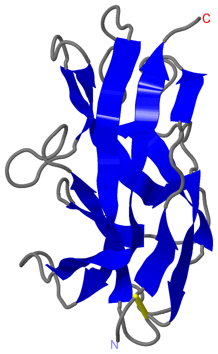
SS Bonds (1, 1)
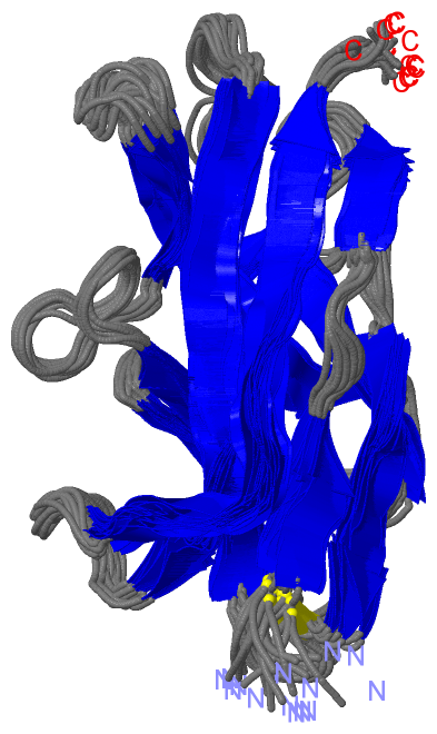
NMR Structure
|
||||||||
Cis Peptide Bonds (0, 0)| (no "Cis Peptide Bond" information available for 1RXL) |
SAPs(SNPs)/Variants (2, 2)
NMR Structure (2, 2)
|
||||||||||||||||||||||||||||||||||||||||||||||||||||||||||||||||||||||||||||||||||||||||||||||||||||||||||||||||||||||||||||||||||||||||||||||
PROSITE Motifs (0, 0)| (no "PROSITE Motif" information available for 1RXL) |
Exons (0, 0)| (no "Exon" information available for 1RXL) |
Sequences/Alignments
NMR StructureChain A from PDB Type:PROTEIN Length:143 aligned with AFAE3_ECOLX | Q57254 from UniProtKB/Swiss-Prot Length:160 Alignment length:143 160 47 57 67 77 87 97 107 117 127 137 147 157 | - - AFAE3_ECOLX 38 EECQVRVGDLTVAKTRGQLTDAAPIGPVTVQALGCNARQVALKADTDNFEQGKFFLISDNNRDKLYVNIRPMDNSAWTTDNGVFYKNDVGSWGGTIGIYVDGQQTNTPPGNYTLTLTGGYWAK-------------------- - SCOP domains d1rxla_ A: DraA/Afimbrial adhesin Afa-III SCOP domains CATH domains 1rxlA00 A:1-143 Afimbrial adhesin afa-iii. CATH domains Pfam domains Adhesin_Dr-1rxlA01 A:1-123 -------------------- Pfam domains SAPs(SNPs) ---------------------T-------------D----------------------------------------------------------------------------------------------------------- SAPs(SNPs) PROSITE ----------------------------------------------------------------------------------------------------------------------------------------------- PROSITE Transcript ----------------------------------------------------------------------------------------------------------------------------------------------- Transcript 1rxl A 1 EECQVRVGDLTVAKTRGQLTDAAPIGPVTVQALGCNARQVALKADTDNFEQGKFFLISDNNRDKLYVNIRPMDNSAWTTDNGVFYKNDVGSWGGTIGIYVDGQQTNTPPGNYTLTLTGGYWAKDNKQGFTPSGTTGTTKLTVT 143 10 20 30 40 50 60 70 80 90 100 110 120 130 140
|
||||||||||||||||||||
SCOP Domains (1, 1)
NMR Structure
 |
CATH Domains (1, 1)
NMR Structure
|
Pfam Domains (1, 1)
NMR Structure
|
Gene Ontology (1, 1)|
NMR Structure(hide GO term definitions) Chain A (AFAE3_ECOLX | Q57254)
|
||||||||||||
Interactive Views
|
||||||||||||||||||||||||||||||||||||||||||||||||||||||||||||||||||||||||||||||||||||||||||||||||||||||||||||||||||||
Still Images
|
||||||||||||||||
Databases
|
||||||||||||||||||||||||||||||||||||||||||||||||||||||||||||||||||||||||||||||||||||||||||||||||||||||||||||||||||||||||||||||||||||||||||||||||||||||||||||||||
Analysis Tools
|
|||||||||||||||||||||||||||||||||||||||||||||||||||||||||||||
Entries Sharing at Least One Protein Chain (UniProt ID)
Related Entries Specified in the PDB File
|
|