Show PDB file:   
         Plain Text   HTML   (compressed file size)
QuickSearch:   
by PDB,NDB,UniProt,PROSITE Code or Search Term(s)  
(-)Asym./Biol. Unit
(-)Asym./Biol. Unit - sites
collapse expand < >
Image Asym./Biol. Unit
Asym./Biol. Unit  (Jmol Viewer)
Image Asym./Biol. Unit - sites
Asym./Biol. Unit - sites  (Jmol Viewer)

(-) Description

Title :  STRUCTURAL AND THERMODYNAMIC DISSECTION OF SPECIFIC MANNAN RECOGNITION BY A CARBOHYDRATE-BINDING MODULE, TMCBM27
 
Authors :  A. B. Boraston, T. J. Revett, C. M. Boraston, D. Nurizzo, G. J. Davies
Date :  07 Apr 03  (Deposition) - 17 Apr 03  (Release) - 24 Feb 09  (Revision)
Method :  X-RAY DIFFRACTION
Resolution :  1.60
Chains :  Asym./Biol. Unit :  A
Keywords :  Hydrolase/Carbohydrate Binding, Mannan Binding, Carbohydrate Binding Module, Polysaccharide Degradation, Hydrolase (Keyword Search: [Gene Ontology, PubMed, Web (Google))
 
Reference :  A. B. Boraston, T. J. Revett, C. M. Boraston, D. Nurizzo, G. J. Davies
Structural And Thermodynamic Dissection Of Specific Mannan Recognition By A Carbohydrate Binding Module, Tmcbm27
Structure V. 11 665 2003
PubMed-ID: 12791255  |  Reference-DOI: 10.1016/S0969-2126(03)00100-X

(-) Compounds

Molecule 1 - BETA-MANNOSIDASE
    ChainsA
    EngineeredYES
    Expression SystemESCHERICHIA COLI
    Expression System PlasmidPET 28-TMCBM27
    Expression System StrainBL21
    Expression System Taxid511693
    Expression System VectorPET 28
    FragmentCARBOHYDRATE-BINDING MODULE, RESIDUES 505-680
    Organism ScientificTHERMOTOGA MARITIMA
    Organism Taxid243274
    StrainMSB8
    SynonymTMCBM27

 Structural Features

(-) Chains, Units

  1
Asymmetric/Biological Unit A

Summary Information (see also Sequences/Alignments below)
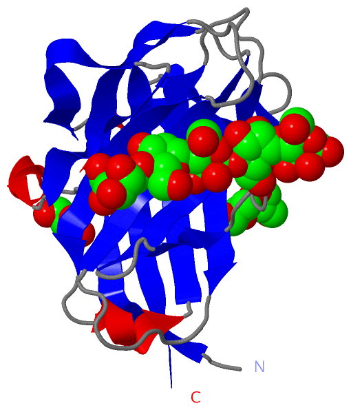
(-) Ligands, Modified Residues, Ions  (3, 8)

Asymmetric/Biological Unit (3, 8)
No.NameCountTypeFull Name
1BMA5Ligand/IonBETA-D-MANNOSE
2CA1Ligand/IonCALCIUM ION
3GOL2Ligand/IonGLYCEROL

(-) Sites  (8, 8)

Asymmetric Unit (8, 8)
No.NameEvidenceResiduesDescription
1AC1SOFTWAREGLU A:31 , LYS A:53 , PRO A:55 , LYS A:57 , ASP A:59 , ARG A:95 , BMA A:1175BINDING SITE FOR RESIDUE BMA A1174
2AC2SOFTWAREGLU A:31 , ASP A:59 , ARG A:95 , ASP A:110 , BMA A:1174 , BMA A:1176BINDING SITE FOR RESIDUE BMA A1175
3AC3SOFTWARETRP A:28 , TRP A:60 , GLU A:62 , LYS A:106 , BMA A:1175 , BMA A:1177 , HOH A:2060 , HOH A:2099 , HOH A:2100BINDING SITE FOR RESIDUE BMA A1176
4AC4SOFTWARETRP A:28 , GLU A:62 , ARG A:64 , LYS A:106 , BMA A:1176 , BMA A:1178 , HOH A:2060 , HOH A:2101BINDING SITE FOR RESIDUE BMA A1177
5AC5SOFTWARESER A:25 , ARG A:64 , TRP A:104 , BMA A:1177 , HOH A:2021 , HOH A:2102 , HOH A:2103 , HOH A:2104 , HOH A:2105BINDING SITE FOR RESIDUE BMA A1178
6AC6SOFTWAREASP A:12 , GLY A:44 , GLY A:46 , ASP A:165 , HOH A:2004 , HOH A:2094BINDING SITE FOR RESIDUE CA A1181
7AC7SOFTWAREGLU A:10 , VAL A:11 , ASP A:12 , PHE A:13 , SER A:14 , SER A:15 , GLU A:18 , HOH A:2106BINDING SITE FOR RESIDUE GOL A1179
8AC8SOFTWAREARG A:4 , SER A:15 , GLU A:17 , VAL A:87 , GLU A:88 , GLU A:120 , ARG A:131 , HIS A:133 , THR A:173 , HOH A:2107BINDING SITE FOR RESIDUE GOL A1180

(-) SS Bonds  (0, 0)

(no "SS Bond" information available for 1OF4)

(-) Cis Peptide Bonds  (2, 2)

Asymmetric/Biological Unit
No.Residues
1Ser A:34 -Pro A:35
2Asn A:101 -Pro A:102

 Sequence-Structure Mapping

(-) SAPs(SNPs)/Variants  (0, 0)

(no "SAP(SNP)/Variant" information available for 1OF4)

(-) PROSITE Motifs  (0, 0)

(no "PROSITE Motif" information available for 1OF4)

(-) Exons   (0, 0)

(no "Exon" information available for 1OF4)

(-) Sequences/Alignments

Asymmetric/Biological Unit
   Reformat: Number of residues per line =  ('0' or empty: single-line sequence representation)
  Number of residues per labelling interval =   
  UniProt sequence: complete  aligned part    
   Show mapping: SCOP domains CATH domains Pfam domains Secondary structure (by author)
SAPs(SNPs) PROSITE motifs Exons
(details for a mapped element are shown in a popup box when the mouse pointer rests over it)
Chain A from PDB  Type:PROTEIN  Length:171
 aligned with Q9RIK9_THEMT | Q9RIK9 from UniProtKB/TrEMBL  Length:680

    Alignment length:171
                                   516       526       536       546       556       566       576       586       596       606       616       626       636       646       656       666       676 
         Q9RIK9_THEMT   507 ARYVLAEEVDFSSPEEVKNWWNSGTWQAEFGSPDIEWNGEVGNGALQLNVKLPGKSDWEEVRVARKFERLSECEILEYDIYIPNVEGLKGRLRPYAVLNPGWVKIGLDMNNANVESAEIITFGGKEYRRFHVRIEFDRTAGVKELHIGVVGDHLRYDGPIFIDNVRLYKRT 677
               SCOP domains d1of4a_ A: Beta-mannosidase, C-terminal domain                                                                                                                              SCOP domains
               CATH domains 1of4A00 A:3-173 Galactose-binding domain-like                                                                                                                               CATH domains
               Pfam domains --------------------------------------------------------------------------------------------------------------------------------------------------------------------------- Pfam domains
         Sec.struct. author ..eeeeeee...hhhhhh.eeeeeee..eeeeeeeeee.hhhhheeeeeeee......eeeeeeee..hhhhh.eeeeeeeee.......ee..eeeee...eee......ee....eeeee..eeeeeeeeeee........eeeeeeeee...eeeeeeeeeeeeeee. Sec.struct. author
                 SAPs(SNPs) --------------------------------------------------------------------------------------------------------------------------------------------------------------------------- SAPs(SNPs)
                    PROSITE --------------------------------------------------------------------------------------------------------------------------------------------------------------------------- PROSITE
                 Transcript --------------------------------------------------------------------------------------------------------------------------------------------------------------------------- Transcript
                 1of4 A   3 ARYVLAEEVDFSSPEEVKNWWNSGTWQAEFGSPDIEWNGEVGNGALQLNVKLPGKSDWEEVRVARKFERLSECEILEYDIYIPNVEGLKGRLRPYAVLNPGWVKIGLDMNNANVESAEIITFGGKEYRRFHVRIEFDRTAGVKELHIGVVGDHLRYDGPIFIDNVRLYKRT 173
                                    12        22        32        42        52        62        72        82        92       102       112       122       132       142       152       162       172 

   Legend:   → Mismatch (orange background)
  - → Gap (green background, '-', border residues have a numbering label)
    → Modified Residue (blue background, lower-case, 'x' indicates undefined single-letter code, labelled with number + name)
  x → Chemical Group (purple background, 'x', labelled with number + name, e.g. ACE or NH2)
  extra numbering lines below/above indicate numbering irregularities and modified residue names etc., number ends below/above '|'

 Classification and Annotation

(-) SCOP Domains  (1, 1)

Asymmetric/Biological Unit

(-) CATH Domains  (1, 1)

Asymmetric/Biological Unit
(-)
Class: Mainly Beta (13760)

(-) Pfam Domains  (0, 0)

(no "Pfam Domain" information available for 1OF4)

(-) Gene Ontology  (6, 6)

Asymmetric/Biological Unit(hide GO term definitions)
Chain A   (Q9RIK9_THEMT | Q9RIK9)
molecular function
    GO:0016787    hydrolase activity    Catalysis of the hydrolysis of various bonds, e.g. C-O, C-N, C-C, phosphoric anhydride bonds, etc. Hydrolase is the systematic name for any enzyme of EC class 3.
    GO:0016798    hydrolase activity, acting on glycosyl bonds    Catalysis of the hydrolysis of any glycosyl bond.
    GO:0004553    hydrolase activity, hydrolyzing O-glycosyl compounds    Catalysis of the hydrolysis of any O-glycosyl bond.
    GO:0046872    metal ion binding    Interacting selectively and non-covalently with any metal ion.
biological process
    GO:0005975    carbohydrate metabolic process    The chemical reactions and pathways involving carbohydrates, any of a group of organic compounds based of the general formula Cx(H2O)y. Includes the formation of carbohydrate derivatives by the addition of a carbohydrate residue to another molecule.
    GO:0008152    metabolic process    The chemical reactions and pathways, including anabolism and catabolism, by which living organisms transform chemical substances. Metabolic processes typically transform small molecules, but also include macromolecular processes such as DNA repair and replication, and protein synthesis and degradation.

 Visualization

(-) Interactive Views

Asymmetric/Biological Unit
  Complete Structure
    Jena3D(integrated viewing of ligand, site, SAP, PROSITE, SCOP information)
    WebMol | AstexViewer[tm]@PDBe
(Java Applets, require no local installation except for Java; loading may be slow)
    STRAP
(Java WebStart application, automatic local installation, requires Java; full application with system access!)
    RasMol
(require local installation)
    Molscript (VRML)
(requires installation of a VRML viewer; select preferred view via VRML and generate a mono or stereo PDF format file)
 
  Ligands, Modified Residues, Ions
    BMA  [ RasMol | Jena3D ]  +environment [ RasMol | Jena3D ]
    CA  [ RasMol | Jena3D ]  +environment [ RasMol | Jena3D ]
    GOL  [ RasMol | Jena3D ]  +environment [ RasMol | Jena3D ]
 
  Sites
    AC1  [ RasMol ]  +environment [ RasMol ]
    AC2  [ RasMol ]  +environment [ RasMol ]
    AC3  [ RasMol ]  +environment [ RasMol ]
    AC4  [ RasMol ]  +environment [ RasMol ]
    AC5  [ RasMol ]  +environment [ RasMol ]
    AC6  [ RasMol ]  +environment [ RasMol ]
    AC7  [ RasMol ]  +environment [ RasMol ]
    AC8  [ RasMol ]  +environment [ RasMol ]
 
  Cis Peptide Bonds
    Asn A:101 - Pro A:102   [ RasMol ]  
    Ser A:34 - Pro A:35   [ RasMol ]  
 

(-) Still Images

Jmol
  protein: cartoon or spacefill or dots and stick; nucleic acid: cartoon and stick; ligands: spacefill; active site: stick
Molscript
  protein, nucleic acid: cartoon; ligands: spacefill; active site: ball and stick

 Databases and Analysis Tools

(-) Databases

Access by PDB/NDB ID
  1of4
    Family and Domain InformationProDom | SYSTERS
    General Structural InformationGlycoscienceDB | MMDB | NDB | OCA | PDB | PDBe | PDBj | PDBsum | PDBWiki | PQS | PROTEOPEDIA
    Orientation in MembranesOPM
    Protein SurfaceSURFACE
    Secondary StructureDSSP (structure derived) | HSSP (homology derived)
    Structural GenomicsGeneCensus
    Structural NeighboursCE | VAST
    Structure ClassificationCATH | Dali | SCOP
    Validation and Original DataBMRB Data View | BMRB Restraints Grid | EDS | PROCHECK | RECOORD | WHAT_CHECK
 
Access by UniProt ID/Accession number
  Q9RIK9_THEMT | Q9RIK9
    Comparative Protein Structure ModelsModBase
    Genomic InformationEnsembl
    Protein-protein InteractionDIP
    Sequence, Family and Domain InformationInterPro | Pfam | SMART | UniProtKB/TrEMBL
 
Access by Enzyme Classificator   (EC Number)
  (no 'Enzyme Classificator' available)
    General Enzyme InformationBRENDA | EC-PDB | Enzyme | IntEnz
    PathwayKEGG | MetaCyc
 
Access by Disease Identifier   (MIM ID)
  (no 'MIM ID' available)
    Disease InformationOMIM
 
Access by GenAge ID
  (no 'GenAge ID' available)
    Age Related InformationGenAge

(-) Analysis Tools

Access by PDB/NDB ID
    Domain InformationXDom
    Interatomic Contacts of Structural UnitsCSU
    Ligand-protein ContactsLPC
    Protein CavitiescastP
    Sequence and Secondary StructurePDBCartoon
    Structure AlignmentSTRAP(Java WebStart application, automatic local installation, requires Java; full application with system access!)
    Structure and Sequence BrowserSTING
 
Access by UniProt ID/Accession number
  Q9RIK9_THEMT | Q9RIK9
    Protein Disorder PredictionDisEMBL | FoldIndex | GLOBPLOT (for more information see DisProt)

 Related Entries

(-) Entries Sharing at Least One Protein Chain (UniProt ID)

UniProtKB/TrEMBL
        Q9RIK9_THEMT | Q9RIK91of3 1oh4

(-) Related Entries Specified in the PDB File

1of3 STRUCTURAL AND THERMODYNAMIC DISSECTION OF SPECIFIC MANNAN RECOGNITION BY A CARBOHYDRATE -BINDING MODULE, TMCBM27