|
|
|
|
 Description
|
|
Compounds
|
||||||||||||||||||||||||||||||||||||||||||||||||||||
Chains, Units
Summary Information (see also Sequences/Alignments below) |
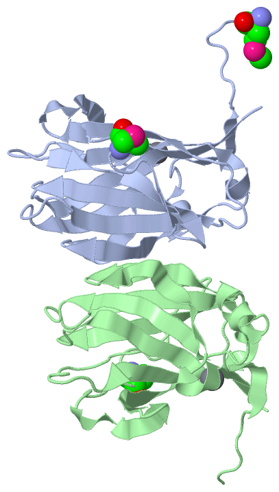
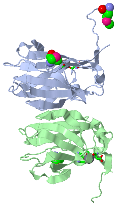
Ligands, Modified Residues, Ions (2, 5)| Asymmetric Unit (2, 5) Biological Unit 1 (1, 2) Biological Unit 2 (1, 1) |
Sites (2, 2)
Asymmetric Unit (2, 2)
|
SS Bonds (0, 0)| (no "SS Bond" information available for 1OF3) |
Cis Peptide Bonds (4, 4)
Asymmetric Unit
|
||||||||||||||||||||
SAPs(SNPs)/Variants (0, 0)| (no "SAP(SNP)/Variant" information available for 1OF3) |
PROSITE Motifs (0, 0)| (no "PROSITE Motif" information available for 1OF3) |
Exons (0, 0)| (no "Exon" information available for 1OF3) |
Sequences/Alignments
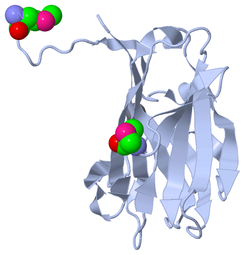
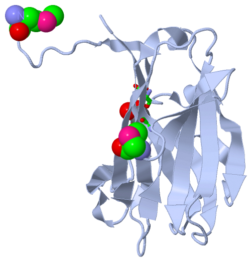
Asymmetric UnitChain A from PDB Type:PROTEIN Length:176 aligned with Q9RIK9_THEMT | Q9RIK9 from UniProtKB/TrEMBL Length:680 Alignment length:176 511 521 531 541 551 561 571 581 591 601 611 621 631 641 651 661 671 Q9RIK9_THEMT 502 KVVNEARYVLAEEVDFSSPEEVKNWWNSGTWQAEFGSPDIEWNGEVGNGALQLNVKLPGKSDWEEVRVARKFERLSECEILEYDIYIPNVEGLKGRLRPYAVLNPGWVKIGLDMNNANVESAEIITFGGKEYRRFHVRIEFDRTAGVKELHIGVVGDHLRYDGPIFIDNVRLYKRT 677 SCOP domains d1of3a_ A: Beta-mannosidase, C-terminal domain SCOP domains CATH domains -1of3A00 A:-1-173 Galactose-binding domain-like CATH domains Pfam domains -------------------------------------------------------------------------------------------------------------------------------------------------------------------------------- Pfam domains SAPs(SNPs) -------------------------------------------------------------------------------------------------------------------------------------------------------------------------------- SAPs(SNPs) PROSITE -------------------------------------------------------------------------------------------------------------------------------------------------------------------------------- PROSITE Transcript -------------------------------------------------------------------------------------------------------------------------------------------------------------------------------- Transcript 1of3 A -2 mASNEARYVLAEEVDFSSPEEVKNWWNSGTWQAEFGSPDIEWNGEVGNGALQLNVKLPGKSDWEEVRVARKFERLSECEILEYDIYIPNVEGLKGRLRPYAVLNPGWVKIGLDmNNANVESAEIITFGGKEYRRFHVRIEFDRTAGVKELHIGVVGDHLRYDGPIFIDNVRLYKRT 173 | 7 17 27 37 47 57 67 77 87 97 107 | 117 127 137 147 157 167 | 111-MSE -2-MSE Chain B from PDB Type:PROTEIN Length:174 aligned with Q9RIK9_THEMT | Q9RIK9 from UniProtKB/TrEMBL Length:680 Alignment length:174 513 523 533 543 553 563 573 583 593 603 613 623 633 643 653 663 673 Q9RIK9_THEMT 504 VNEARYVLAEEVDFSSPEEVKNWWNSGTWQAEFGSPDIEWNGEVGNGALQLNVKLPGKSDWEEVRVARKFERLSECEILEYDIYIPNVEGLKGRLRPYAVLNPGWVKIGLDMNNANVESAEIITFGGKEYRRFHVRIEFDRTAGVKELHIGVVGDHLRYDGPIFIDNVRLYKRT 677 SCOP domains d1of3b_ B: Beta-mannosidase, C-terminal domain SCOP domains CATH domains 1of3B00 B:0-173 Galactose-binding domain-like CATH domains Pfam domains ------------------------------------------------------------------------------------------------------------------------------------------------------------------------------ Pfam domains SAPs(SNPs) ------------------------------------------------------------------------------------------------------------------------------------------------------------------------------ SAPs(SNPs) PROSITE ------------------------------------------------------------------------------------------------------------------------------------------------------------------------------ PROSITE Transcript ------------------------------------------------------------------------------------------------------------------------------------------------------------------------------ Transcript 1of3 B 0 SNEARYVLAEEVDFSSPEEVKNWWNSGTWQAEFGSPDIEWNGEVGNGALQLNVKLPGKSDWEEVRVARKFERLSECEILEYDIYIPNVEGLKGRLRPYAVLNPGWVKIGLDmNNANVESAEIITFGGKEYRRFHVRIEFDRTAGVKELHIGVVGDHLRYDGPIFIDNVRLYKRT 173 9 19 29 39 49 59 69 79 89 99 109 | 119 129 139 149 159 169 111-MSE
|
||||||||||||||||||||
SCOP Domains (1, 2)
Asymmetric Unit
 |
CATH Domains (1, 2)
Asymmetric Unit
|
Pfam Domains (0, 0)| (no "Pfam Domain" information available for 1OF3) |
Gene Ontology (6, 6)|
Asymmetric Unit(hide GO term definitions) Chain A,B (Q9RIK9_THEMT | Q9RIK9)
|
||||||||||||||||||||||||||||||||||||||||||||||||
Interactive Views
|
|||||||||||||||||||||||||||||||||||||||||||||||||||||||||||||||||||||||||||||||||||||||||||||||||||||||||||||||||||||||||||||||||||||||||||||||||||||||||||||||||||||||||||||||||
Still Images
|
||||||||||||||||
Databases
|
||||||||||||||||||||||||||||||||||||||||||||||||||||||||||||||||||||||||||||||||||||||||||||||||||||||||||||||||||||||||||||||||||||||||||||||||||||||||||||||||
Analysis Tools
|
|||||||||||||||||||||||||||||||||||||||||||||||||||||||||||||
Entries Sharing at Least One Protein Chain (UniProt ID)
Related Entries Specified in the PDB File
|
|