| molecular function |
|---|
| | GO:0001848 | | complement binding | | Interacting selectively and non-covalently with any component or product of the complement cascade. |
| | GO:0032403 | | protein complex binding | | Interacting selectively and non-covalently with any protein complex (a complex of two or more proteins that may include other nonprotein molecules). |
| | GO:0019841 | | retinol binding | | Interacting selectively and non-covalently with retinol, vitamin A1, 2,6,6-trimethyl-1-(9'-hydroxy-3',7'-dimethylnona-1',3',5',7'-tetraenyl)cyclohex-1-ene, one of the three components that makes up vitamin A. Retinol is an intermediate in the vision cycle and it also plays a role in growth and differentiation. |
| | GO:0036094 | | small molecule binding | | Interacting selectively and non-covalently with a small molecule, any low molecular weight, monomeric, non-encoded molecule. |
| biological process |
|---|
| | GO:0006957 | | complement activation, alternative pathway | | Any process involved in the activation of any of the steps of the alternative pathway of the complement cascade which allows for the direct killing of microbes and the regulation of other immune processes. |
| | GO:0006958 | | complement activation, classical pathway | | Any process involved in the activation of any of the steps of the classical pathway of the complement cascade which allows for the direct killing of microbes, the disposal of immune complexes, and the regulation of other immune processes. |
| | GO:0019835 | | cytolysis | | The rupture of cell membranes and the loss of cytoplasm. |
| | GO:0002376 | | immune system process | | Any process involved in the development or functioning of the immune system, an organismal system for calibrated responses to potential internal or invasive threats. |
| | GO:0045087 | | innate immune response | | Innate immune responses are defense responses mediated by germline encoded components that directly recognize components of potential pathogens. |
| | GO:0030449 | | regulation of complement activation | | Any process that modulates the frequency, rate or extent of complement activation. |
| cellular component |
|---|
| | GO:0072562 | | blood microparticle | | A phospholipid microvesicle that is derived from any of several cell types, such as platelets, blood cells, endothelial cells, or others, and contains membrane receptors as well as other proteins characteristic of the parental cell. Microparticles are heterogeneous in size, and are characterized as microvesicles free of nucleic acids. |
| | GO:0070062 | | extracellular exosome | | A vesicle that is released into the extracellular region by fusion of the limiting endosomal membrane of a multivesicular body with the plasma membrane. Extracellular exosomes, also simply called exosomes, have a diameter of about 40-100 nm. |
| | GO:0005576 | | extracellular region | | The space external to the outermost structure of a cell. For cells without external protective or external encapsulating structures this refers to space outside of the plasma membrane. This term covers the host cell environment outside an intracellular parasite. |
| | GO:0005615 | | extracellular space | | That part of a multicellular organism outside the cells proper, usually taken to be outside the plasma membranes, and occupied by fluid. |
| | GO:0005579 | | membrane attack complex | | A protein complex produced by sequentially activated components of the complement cascade inserted into a target cell membrane and forming a pore leading to cell lysis via ion and water flow. |
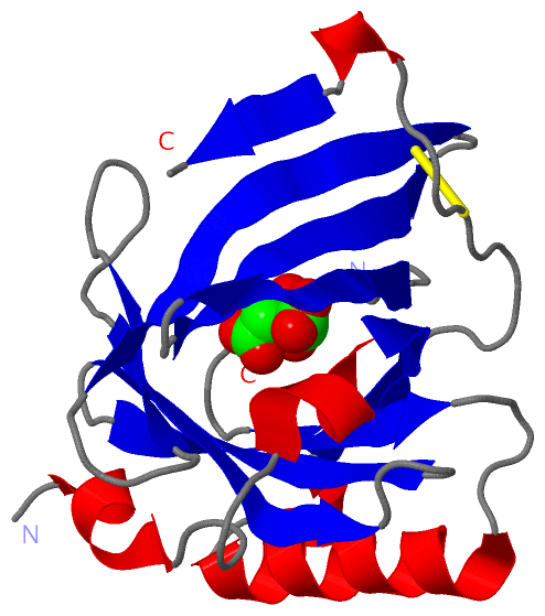
 Description
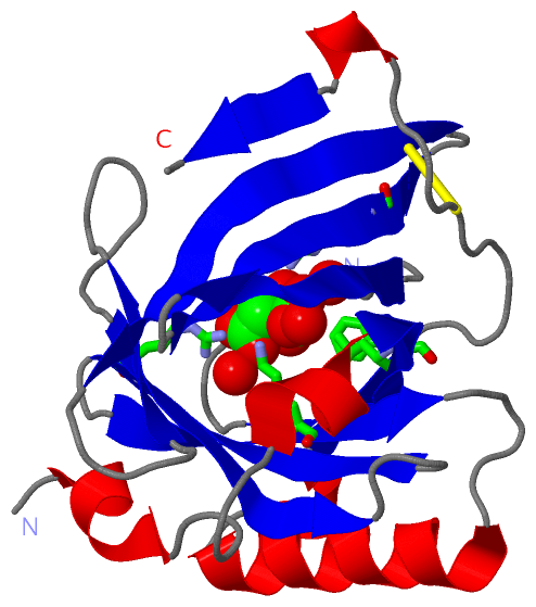