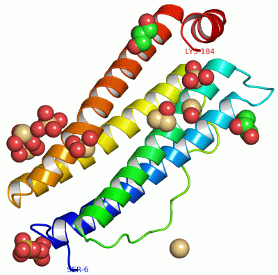
Asymmetric Unit (13, 13)
| No. | Name | Evidence | Residues | Description |
|---|
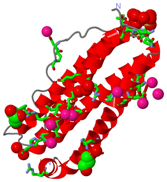
| 01 | AC1 | SOFTWARE | GLN A:10 , ASN A:11 , HOH A:204 , HOH A:209 , HOH A:310 , HOH A:372 | BINDING SITE FOR RESIDUE SO4 A 187 |
| 02 | AC2 | SOFTWARE | SER A:13 , THR A:14 , GLU A:15 , ARG A:124 | BINDING SITE FOR RESIDUE SO4 A 188 |
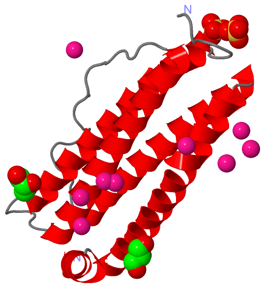
| 03 | AC3 | SOFTWARE | ASP A:84 , GLN A:86 | BINDING SITE FOR RESIDUE CD A 717 |
| 04 | AC4 | SOFTWARE | GLU A:60 , GLU A:64 , HOH A:505 , HOH A:506 , CD A:722 | BINDING SITE FOR RESIDUE CD A 718 |
| 05 | AC5 | SOFTWARE | GLU A:61 , GLU A:64 , HOH A:505 , HOH A:506 , CD A:718 | BINDING SITE FOR RESIDUE CD A 722 |
| 06 | AC6 | SOFTWARE | GLU A:57 , GLU A:60 , HOH A:507 | BINDING SITE FOR RESIDUE CD A 723 |
| 07 | AC7 | SOFTWARE | ASP A:131 , SER A:135 , HOH A:508 , HOH A:509 , HOH A:510 , HOH A:511 , HOH A:512 | BINDING SITE FOR RESIDUE CD A 715 |
| 08 | AC8 | SOFTWARE | GLU A:134 , HOH A:452 , HOH A:513 , HOH A:514 , HOH A:515 , HOH A:520 | BINDING SITE FOR RESIDUE CD A 716 |
| 09 | AC9 | SOFTWARE | HIS A:136 , HOH A:409 , HOH A:516 , HOH A:517 , HOH A:518 | BINDING SITE FOR RESIDUE CD A 719 |
| 10 | BC1 | SOFTWARE | HIS A:118 , CYS A:130 , GLU A:134 , HOH A:435 , HOH A:452 , HOH A:513 , HOH A:515 , HOH A:519 , HOH A:520 , HOH A:521 | BINDING SITE FOR RESIDUE CD A 720 |
| 11 | BC2 | SOFTWARE | HIS A:53 , GLU A:57 , HOH A:522 , HOH A:523 , HOH A:524 | BINDING SITE FOR RESIDUE CD A 721 |
| 12 | BC3 | SOFTWARE | GLU A:49 , LYS A:147 , ASN A:150 , ARG A:180 , HOH A:239 , HOH A:348 , HOH A:349 , HOH A:380 | BINDING SITE FOR RESIDUE GOL A 724 |
| 13 | BC4 | SOFTWARE | PHE A:39 , ARG A:56 , GLU A:67 , ARG A:68 , HOH A:229 , HOH A:250 , HOH A:442 , HOH A:443 | BINDING SITE FOR RESIDUE GOL A 189 |
|
 Description