|
|
|
|
 Description
|
|
Compounds
|
||||||||||||||||||||||||||||||||||||||||||||||||||||||||
Chains, Units
Summary Information (see also Sequences/Alignments below) |
Ligands, Modified Residues, Ions (2, 10)| Asymmetric Unit (2, 10) Biological Unit 1 (1, 24) |
Sites (10, 10)
Asymmetric Unit (10, 10)
|
SS Bonds (0, 0)| (no "SS Bond" information available for 1H96) |
Cis Peptide Bonds (0, 0)| (no "Cis Peptide Bond" information available for 1H96) |
SAPs(SNPs)/Variants (0, 0)| (no "SAP(SNP)/Variant" information available for 1H96) |
PROSITE Motifs (3, 3)
Asymmetric Unit (3, 3)
|
||||||||||||||||||||||||||||||||||||||||||||||||||||||||||||||||||||||||||||||||
Exons (0, 0)| (no "Exon" information available for 1H96) |
Sequences/Alignments
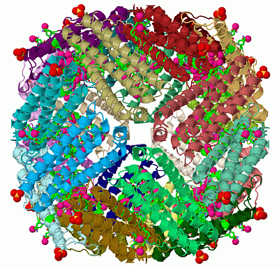
Asymmetric UnitChain A from PDB Type:PROTEIN Length:169 aligned with FRIL1_MOUSE | P29391 from UniProtKB/Swiss-Prot Length:183 Alignment length:180 11 21 31 41 51 61 71 81 91 101 111 121 131 141 151 161 171 181 FRIL1_MOUSE 2 TSQIRQNYSTEVEAAVNRLVNLHLRASYTYLSLGFFFDRDDVALEGVGHFFRELAEEKREGAERLLEFQNDRGGRALFQDVQKPSQDEWGKTQEAMEAALAMEKNLNQALLDLHALGSARTDPHLCDFLESHYLDKEVKLIKKMGNHLTNLRRVAGPQPAQTGAPQGSLGEYLFERLTLK 181 SCOP domains d1h96a_ A: (Apo)ferritin SCOP domains CATH domains 1h96A00 A:1-180 [code=1.20.1260.10, no name defined] CATH domains Pfam domains ------------------------------------------------------------------------------------------------------------------------------------------------------------------------------------ Pfam domains SAPs(SNPs) ------------------------------------------------------------------------------------------------------------------------------------------------------------------------------------ SAPs(SNPs) PROSITE (1) -----FERRITIN_LIKE PDB: A:6-155 UniProt: 7-156 ------------------------- PROSITE (1) PROSITE (2) --------------------------------------------------------FERRITIN_1 ----------------------------------------------FERRITIN_2 -------------------------------------- PROSITE (2) Transcript ------------------------------------------------------------------------------------------------------------------------------------------------------------------------------------ Transcript 1h96 A 1 TSQIRQNYSTEVEAAVNRLVNLHLRASYTYLSLGFFFDRDDVALEGVGHFFRELAEEKREGAERLLEFQNDRGGRALFQDVQKPSQDEWGKTQEAMEAALAMEKNLNQALLDLHALGSARADPHLCDFLESHYLDKEVKLIKKMGNHLTNLRRVAG-----------SLGEYLFERLTLK 180 10 20 30 40 50 60 70 80 90 100 110 120 130 140 150 | - 170 180 156 168
|
||||||||||||||||||||
SCOP Domains (1, 1)
Asymmetric Unit
 |
CATH Domains (1, 1)
Asymmetric Unit
|
Pfam Domains (0, 0)| (no "Pfam Domain" information available for 1H96) |
Gene Ontology (13, 13)|
Asymmetric Unit(hide GO term definitions) Chain A (FRIL1_MOUSE | P29391)
|
||||||||||||||||||||||||||||||||||||||||||||||||||||||||||||||||||||||||||||||||||||||||||||||||
Interactive Views
|
||||||||||||||||||||||||||||||||||||||||||||||||||||||||||||||||||||||||||||||||||||||||||||||||||||||||||||||||||||||||||||||||||||||||||||||||||||||||||||||||||||||||||||||||||||||||||||||||||||||||||||||
Still Images
|
||||||||||||||||
Databases
|
||||||||||||||||||||||||||||||||||||||||||||||||||||||||||||||||||||||||||||||||||||||||||||||||||||||||||||||||||||||||||||||||||||||||||||||||||||||||||||||||
Analysis Tools
|
|||||||||||||||||||||||||||||||||||||||||||||||||||||||||||||
Entries Sharing at Least One Protein Chain (UniProt ID)
Related Entries Specified in the PDB File
|
|