Show PDB file:   
         Plain Text   HTML   (compressed file size)
QuickSearch:   
by PDB,NDB,UniProt,PROSITE Code or Search Term(s)  
(-)Asym./Biol. Unit - manually
(-)Asym./Biol. Unit
(-)Asym./Biol. Unit - sites
collapse expand < >
Image Asym./Biol. Unit - manually
Asym./Biol. Unit - manually  (Jmol Viewer)
Image Asym./Biol. Unit
Asym./Biol. Unit  (Jmol Viewer)
Image Asym./Biol. Unit - sites
Asym./Biol. Unit - sites  (Jmol Viewer)

(-) Description

Title :  CRYSTAL STRUCTURE OF THE ANTI-ERBB2 FAB2C4
 
Authors :  F. F. Vajdos, C. W. Adams, T. N. Breece, L. G. Presta, A. M. De Vos, S. S. Sidhu
Date :  15 Mar 02  (Deposition) - 10 Jul 02  (Release) - 24 Feb 09  (Revision)
Method :  X-RAY DIFFRACTION
Resolution :  1.80
Chains :  Asym./Biol. Unit :  H,L
Keywords :  Ig Domain, Fab Fragment, Immune System (Keyword Search: [Gene Ontology, PubMed, Web (Google))
 
Reference :  F. F. Vajdos, C. W. Adams, T. N. Breece, L. G. Presta, A. M. De Vos, S. S. Sidhu
Comprehensive Functional Maps Of The Antigen-Binding Site Of An Anti-Erbb2 Antibody Obtained With Shotgun Scanning Mutagenesis.
J. Mol. Biol. V. 320 415 2002
PubMed-ID: 12079396  |  Reference-DOI: 10.1016/S0022-2836(02)00264-4
(for further references see the PDB file header)

(-) Compounds

Molecule 1
    ChainsL
    EngineeredYES
    Expression SystemESCHERICHIA COLI
    Expression System Taxid562
    FragmentLIGHT CHAIN (RESIDUES 1-214)
    Organism CommonHUMAN
    Organism ScientificHOMO SAPIENS
    Organism Taxid9606
    Other DetailsTHE CHIMERA CONSISTS OF THE HUMAN IG AND THE ORIGINAL MURINE SEQUENCE
 
Molecule 2
    ChainsH
    EngineeredYES
    Expression SystemESCHERICHIA COLI
    Expression System Taxid562
    FragmentHEAVY CHAIN (RESIDUES 1-216)
    Organism CommonHUMAN
    Organism ScientificHOMO SAPIENS
    Organism Taxid9606
    Other DetailsTHE CHIMERA CONSISTS OF THE HUMAN IG AND THE ORIGINAL MURINE SEQUENCE

 Structural Features

(-) Chains, Units

  12
Asymmetric/Biological Unit HL

Summary Information (see also Sequences/Alignments below)

(-) Ligands, Modified Residues, Ions  (1, 2)

Asymmetric/Biological Unit (1, 2)
No.NameCountTypeFull Name
1SO42Ligand/IonSULFATE ION

(-) Sites  (2, 2)

Asymmetric Unit (2, 2)
No.NameEvidenceResiduesDescription
1AC1SOFTWAREARG L:54 , THR L:56 , GLY L:57 , HOH L:564 , HOH L:609 , HOH L:615BINDING SITE FOR RESIDUE SO4 L 401
2AC2SOFTWAREPRO L:59 , SER L:60 , HOH L:488 , HOH L:583BINDING SITE FOR RESIDUE SO4 L 402

(-) SS Bonds  (5, 5)

Asymmetric/Biological Unit
No.Residues
1H:22 -H:92
2H:140 -H:196
3L:23 -L:88
4L:134 -L:194
5L:214 -H:216

(-) Cis Peptide Bonds  (5, 5)

Asymmetric/Biological Unit
No.Residues
1Ser L:7 -Pro L:8
2Tyr L:94 -Pro L:95
3Tyr L:140 -Pro L:141
4Phe H:146 -Pro H:147
5Glu H:148 -Pro H:149

 Sequence-Structure Mapping

(-) SAPs(SNPs)/Variants  (0, 0)

(no "SAP(SNP)/Variant" information available for 1L7I)

(-) PROSITE Motifs  (0, 0)

(no "PROSITE Motif" information available for 1L7I)

(-) Exons   (0, 0)

(no "Exon" information available for 1L7I)

(-) Sequences/Alignments

Asymmetric/Biological Unit
   Reformat: Number of residues per line =  ('0' or empty: single-line sequence representation)
  Number of residues per labelling interval =   
  UniProt sequence: complete  aligned part    
   Show mapping: SCOP domains CATH domains Pfam domains Secondary structure (by author)
SAPs(SNPs) PROSITE motifs Exons
(details for a mapped element are shown in a popup box when the mouse pointer rests over it)
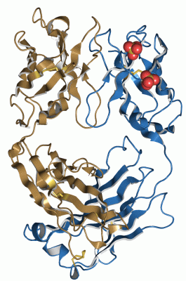
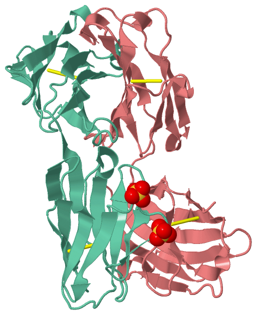
Chain H from PDB  Type:PROTEIN  Length:222
                                                                                                                                                                                                                                                              
               SCOP domains d1l7ih1 H:1-113 Immunoglobulin heavy chain variable domain, VH                                                         d1l7ih2 H:114-216 Immunoglobulin heavy chain gamma constant domain 1, CH1-gamma                         SCOP domains
               CATH domains 1l7iH01 H:1-113 Immunoglobulins                                                                                        1l7iH02 H:114-213 Immunoglobulins                                                                   --- CATH domains
               Pfam domains ------------------------------------------------------------------------------------------------------------------------------------------------------------------------------------------------------------------------------ Pfam domains
         Sec.struct. author ..eeeee...ee.....eeeeeeee..hhhhheeeeeee......eeeeee......eee.hhhh..eeeeee....eeeeee...hhhhheeeeeeee........ee...eeeee........eeeee..hhh.ee..eeeeeeeeeee.....eeee.hhh....eee...ee.....eeeeeeeeee.hhh.....eeeeeehhhheeeeee...... Sec.struct. author
                 SAPs(SNPs) ------------------------------------------------------------------------------------------------------------------------------------------------------------------------------------------------------------------------------ SAPs(SNPs)
                    PROSITE ------------------------------------------------------------------------------------------------------------------------------------------------------------------------------------------------------------------------------ PROSITE
                 Transcript ------------------------------------------------------------------------------------------------------------------------------------------------------------------------------------------------------------------------------ Transcript
                 1l7i H   1 EVQLVESGGGLVQPGGSLRLSCAASGFTFTDYTMDWVRQAPGKGLEWVADVNPNSGGSIYNQRFKGRFTLSVDRSKNTLYLQMNSLRAEDTAVYYCARNLGPSFYFDYWGQGTLVTVSSASTKGPSVFPLAPSSKSTSGGTAALGCLVKDYFPEPVTVSWNSGALTSGVHTFPAVLQSSGLYSLSSVVTVPSSSLGTQTYICNVNHKPSNTKVDKKVEPKSC 216
                                    10        20        30        40        50  |     59        69        79   |||  86        96   ||  104       114       124       134       144       154       164       174       184       194       204       214  
                                                                              52A                            82A||               99A|                                                                                                                     
                                                                                                              82B|                99B                                                                                                                     
                                                                                                               82C                                                                                                                                        

Chain L from PDB  Type:PROTEIN  Length:214
 aligned with Q7Z3Y4_HUMAN | Q7Z3Y4 from UniProtKB/TrEMBL  Length:236

    Alignment length:214
                                    32        42        52        62        72        82        92       102       112       122       132       142       152       162       172       182       192       202       212       222       232    
         Q7Z3Y4_HUMAN    23 DIQMTQSPSSLSASVGDTVTITCRASQDISNYLAWFQQKPGKAPKSLIYGASSLQSGVQSKFSGSGSGTDFTLTISSLQPEDFATYYCQQYKSYPVTFGQGTKLEIKRTVAAPSVFIFPPSDEQLKSGTASVVCLLNNFYPREAKVQWKVDNALQSGNSQESVTEQDSKDSTYSLSSTLTLSKADYEKHKVYACEVTHQGLSSPVTKSFNRGEC 236
               SCOP domains d1l7il1 L:1-107 Immunoglobulin light chain kappa variable domain, VL-kappa                                 d1l7il2 L:108-214 Immunoglobulin light chain kappa constant domain, CL-kappa                                SCOP domains
               CATH domains 1l7iL01 L:1-108 Immunoglobulins                                                                             1l7iL02 L:109-211 Immunoglobulins                                                                      --- CATH domains
               Pfam domains ---------------------------------------------------------------------------------------------------------------------------------------------------------------------------------------------------------------------- Pfam domains
         Sec.struct. author ...eeee..eeeee....eeeeeee.......eeeeee......eeeee...ee.......eeeeee..eeeeee...hhhhheeeeeee...........eeeeee......eeeee..hhhhhh..eeeeeeeeeee.....eeeeee..ee....eeeee.........eeeeeeeeeehhhhh...eeeeeee.......eeeeee.hhh Sec.struct. author
                 SAPs(SNPs) ---------------------------------------------------------------------------------------------------------------------------------------------------------------------------------------------------------------------- SAPs(SNPs)
                    PROSITE ---------------------------------------------------------------------------------------------------------------------------------------------------------------------------------------------------------------------- PROSITE
                 Transcript ---------------------------------------------------------------------------------------------------------------------------------------------------------------------------------------------------------------------- Transcript
                 1l7i L   1 DIQMTQSPSSLSASVGDRVTITCKASQDVSIGVAWYQQKPGKAPKLLIYSASYRYTGVPSRFSGSGSGTDFTLTISSLQPEDFATYYCQQYYIYPYTFGQGTKVEIKRTVAAPSVFIFPPSDEQLKSGTASVVCLLNNFYPREAKVQWKVDNALQSGNSQESVTEQDSKDSTYSLSSTLTLSKADYEKHKVYACEVTHQGLSSPVTKSFNRGEC 214
                                    10        20        30        40        50        60        70        80        90       100       110       120       130       140       150       160       170       180       190       200       210    

   Legend:   → Mismatch (orange background)
  - → Gap (green background, '-', border residues have a numbering label)
    → Modified Residue (blue background, lower-case, 'x' indicates undefined single-letter code, labelled with number + name)
  x → Chemical Group (purple background, 'x', labelled with number + name, e.g. ACE or NH2)
  extra numbering lines below/above indicate numbering irregularities and modified residue names etc., number ends below/above '|'

 Classification and Annotation

(-) SCOP Domains  (4, 4)

Asymmetric/Biological Unit

(-) CATH Domains  (1, 4)

Asymmetric/Biological Unit
(-)
Class: Mainly Beta (13760)

(-) Pfam Domains  (0, 0)

(no "Pfam Domain" information available for 1L7I)

(-) Gene Ontology  (0, 0)

Asymmetric/Biological Unit(hide GO term definitions)
    (no "Gene Ontology" information available for 1L7I)

 Visualization

(-) Interactive Views

Asymmetric/Biological Unit
  Complete Structure
    Jena3D(integrated viewing of ligand, site, SAP, PROSITE, SCOP information)
    WebMol | AstexViewer[tm]@PDBe
(Java Applets, require no local installation except for Java; loading may be slow)
    STRAP
(Java WebStart application, automatic local installation, requires Java; full application with system access!)
    RasMol
(require local installation)
    Molscript (VRML)
(requires installation of a VRML viewer; select preferred view via VRML and generate a mono or stereo PDF format file)
 
  Ligands, Modified Residues, Ions
    SO4  [ RasMol | Jena3D ]  +environment [ RasMol | Jena3D ]
 
  Sites
    AC1  [ RasMol ]  +environment [ RasMol ]
    AC2  [ RasMol ]  +environment [ RasMol ]
 
  Cis Peptide Bonds
    Glu H:148 - Pro H:149   [ RasMol ]  
    Phe H:146 - Pro H:147   [ RasMol ]  
    Ser L:7 - Pro L:8   [ RasMol ]  
    Tyr L:140 - Pro L:141   [ RasMol ]  
    Tyr L:94 - Pro L:95   [ RasMol ]  
 

(-) Still Images

Jmol
  protein: cartoon or spacefill or dots and stick; nucleic acid: cartoon and stick; ligands: spacefill; active site: stick
Molscript
  protein, nucleic acid: cartoon; ligands: spacefill; active site: ball and stick

 Databases and Analysis Tools

(-) Databases

Access by PDB/NDB ID
  1l7i
    Family and Domain InformationProDom | SYSTERS
    General Structural InformationGlycoscienceDB | MMDB | NDB | OCA | PDB | PDBe | PDBj | PDBsum | PDBWiki | PQS | PROTEOPEDIA
    Orientation in MembranesOPM
    Protein SurfaceSURFACE
    Secondary StructureDSSP (structure derived) | HSSP (homology derived)
    Structural GenomicsGeneCensus
    Structural NeighboursCE | VAST
    Structure ClassificationCATH | Dali | SCOP
    Validation and Original DataBMRB Data View | BMRB Restraints Grid | EDS | PROCHECK | RECOORD | WHAT_CHECK
 
Access by UniProt ID/Accession number
  Q7Z3Y4_HUMAN | Q7Z3Y4
    Comparative Protein Structure ModelsModBase
    Genomic InformationEnsembl
    Protein-protein InteractionDIP
    Sequence, Family and Domain InformationInterPro | Pfam | SMART | UniProtKB/TrEMBL
 
Access by Enzyme Classificator   (EC Number)
  (no 'Enzyme Classificator' available)
    General Enzyme InformationBRENDA | EC-PDB | Enzyme | IntEnz
    PathwayKEGG | MetaCyc
 
Access by Disease Identifier   (MIM ID)
  (no 'MIM ID' available)
    Disease InformationOMIM
 
Access by GenAge ID
  (no 'GenAge ID' available)
    Age Related InformationGenAge

(-) Analysis Tools

Access by PDB/NDB ID
    Domain InformationXDom
    Interatomic Contacts of Structural UnitsCSU
    Ligand-protein ContactsLPC
    Protein CavitiescastP
    Sequence and Secondary StructurePDBCartoon
    Structure AlignmentSTRAP(Java WebStart application, automatic local installation, requires Java; full application with system access!)
    Structure and Sequence BrowserSTING
 
Access by UniProt ID/Accession number
  Q7Z3Y4_HUMAN | Q7Z3Y4
    Protein Disorder PredictionDisEMBL | FoldIndex | GLOBPLOT (for more information see DisProt)

 Related Entries

(-) Entries Sharing at Least One Protein Chain (UniProt ID)

UniProtKB/TrEMBL
        Q7Z3Y4_HUMAN | Q7Z3Y41axs 1pg7 1s78 2jix 4ogx 4ogy 4pub

(-) Related Entries Specified in the PDB File

(no "Related Entries Specified in the PDB File" available for 1L7I)