Show PDB file:   
         Plain Text   HTML   (compressed file size)
QuickSearch:   
by PDB,NDB,UniProt,PROSITE Code or Search Term(s)  
(-)Asym./Biol. Unit - manually
(-)Asym./Biol. Unit
(-)Asym./Biol. Unit - sites
collapse expand < >
Image Asym./Biol. Unit - manually
Asym./Biol. Unit - manually  (Jmol Viewer)
Image Asym./Biol. Unit
Asym./Biol. Unit  (Jmol Viewer)
Image Asym./Biol. Unit - sites
Asym./Biol. Unit - sites  (Jmol Viewer)

(-) Description

Title :  CRYSTAL STRUCTURE OF AN AMPICILLIN SINGLE CHAIN FV, FORM 1, FREE
 
Authors :  S. Jung, S. Spinelli, B. Schimmele, A. Honegger, L. Pugliese, C. Cambillau, A. Pluckthun
Date :  15 Feb 01  (Deposition) - 17 Oct 01  (Release) - 24 Feb 09  (Revision)
Method :  X-RAY DIFFRACTION
Resolution :  2.44
Chains :  Asym./Biol. Unit :  H,L
Keywords :  Antibody Fv Fragment, Antibiotic (Keyword Search: [Gene Ontology, PubMed, Web (Google))
 
Reference :  J. Burmester, S. Spinelli, L. Pugliese, A. Krebber, A. Honegger, S. Jung, B. Schimmele, C. Cambillau, A. Pluckthun
Selection, Characterization And X-Ray Structure Of Anti-Ampicillin Single-Chain Fv Fragments From Phage-Displayed Murine Antibody Libraries.
J. Mol. Biol. V. 309 671 2001
PubMed-ID: 11397088  |  Reference-DOI: 10.1006/JMBI.2001.4663
(for further references see the PDB file header)

(-) Compounds

Molecule 1 - ANTIBODY FV FRAGMENT
    ChainsL
    EngineeredYES
    Expression SystemESCHERICHIA COLI
    Expression System Taxid562
    Expression System Vector TypePLASMID
    FragmentVL DOMAIN
    Organism CommonHOUSE MOUSE
    Organism ScientificMUS MUSCULUS
    Organism Taxid10090
    Other DetailsPROTEIN SELECTED BY PHAGE DISPLAY
 
Molecule 2 - ANTIBODY FV FRAGMENT
    ChainsH
    EngineeredYES
    Expression SystemESCHERICHIA COLI
    Expression System Taxid562
    Expression System Vector TypePLASMID
    FragmentVH DOMAIN
    Organism CommonHOUSE MOUSE
    Organism ScientificMUS MUSCULUS
    Organism Taxid10090
    Other DetailsPROTEIN SELECTED BY PHAGE DISPLAY

 Structural Features

(-) Chains, Units

  12
Asymmetric/Biological Unit HL

Summary Information (see also Sequences/Alignments below)

(-) Ligands, Modified Residues, Ions  (1, 1)

Asymmetric/Biological Unit (1, 1)
No.NameCountTypeFull Name
1MPD1Ligand/Ion(4S)-2-METHYL-2,4-PENTANEDIOL

(-) Sites  (1, 1)

Asymmetric Unit (1, 1)
No.NameEvidenceResiduesDescription
1AC1SOFTWAREASN H:42 , TRP H:139 , TYR L:44 , GLN L:107 , TYR L:109 , HOH L:436BINDING SITE FOR RESIDUE MPD L 500

(-) SS Bonds  (2, 2)

Asymmetric/Biological Unit
No.Residues
1H:23 -H:106
2L:23 -L:106

(-) Cis Peptide Bonds  (1, 1)

Asymmetric/Biological Unit
No.Residues
1Tyr L:135 -Pro L:136

 Sequence-Structure Mapping

(-) SAPs(SNPs)/Variants  (0, 0)

(no "SAP(SNP)/Variant" information available for 1I3G)

(-) PROSITE Motifs  (0, 0)

(no "PROSITE Motif" information available for 1I3G)

(-) Exons   (0, 0)

(no "Exon" information available for 1I3G)

(-) Sequences/Alignments

Asymmetric/Biological Unit
   Reformat: Number of residues per line =  ('0' or empty: single-line sequence representation)
  Number of residues per labelling interval =   
  UniProt sequence: complete  aligned part    
   Show mapping: SCOP domains CATH domains Pfam domains Secondary structure (by author)
SAPs(SNPs) PROSITE motifs Exons
(details for a mapped element are shown in a popup box when the mouse pointer rests over it)
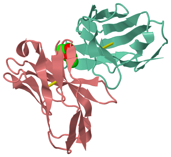
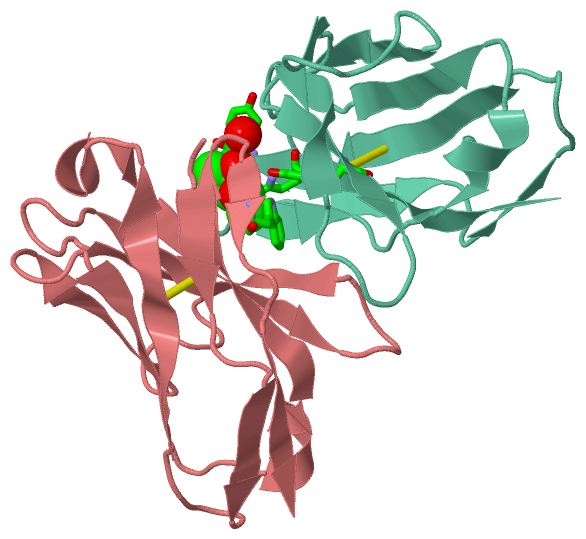
Chain H from PDB  Type:PROTEIN  Length:111
                                                                                                                                               
               SCOP domains d1i3gh_ H: Immunoglobulin heavy chain variable domain, VH                                                       SCOP domains
               CATH domains 1i3gH00 H:1-148 Immunoglobulins                                                                                 CATH domains
               Pfam domains --------------------------------------------------------------------------------------------------------------- Pfam domains
         Sec.struct. author ..eee...eeee.....eeeeeeee..hhhhh.eeeeee.....eeeeeee......eee.......eeeeee....eeeeee...hhhhheeeeeee..ee...eeeee. Sec.struct. author
                 SAPs(SNPs) --------------------------------------------------------------------------------------------------------------- SAPs(SNPs)
                    PROSITE --------------------------------------------------------------------------------------------------------------- PROSITE
                 Transcript --------------------------------------------------------------------------------------------------------------- Transcript
                 1i3g H   1 QVQLQQPGAELVRPGASVKLSCKASGYTFTSYWINWVKQRPGQGLEWIGNIYPSDSYTNYNQKFKDKATLTVDKSSSTAYMQLSSLTSEDSAVYFCARWGYWGQGTLVTVS 148
                                  ||11        21     || 32||      47        57   ||   70        80        90       100       137       147 
                                  7|                27|  33|                    61|                                         109|           
                                   9                 29   39                     65                                          137           

Chain L from PDB  Type:PROTEIN  Length:106
                                                                                                                                          
               SCOP domains d1i3gl_ L: Immunoglobulin light chain kappa variable domain, VL-kappa                                      SCOP domains
               CATH domains 1i3gL00 L:1-147 Immunoglobulins                                                                            CATH domains
               Pfam domains ---------------------------------------------------------------------------------------------------------- Pfam domains
         Sec.struct. author ...eee...eee......eeeeeee.......eeeeee......eeeee...ee.......eeeeee..eeeeee...hhhhh.eeeeee......ee...eeee. Sec.struct. author
                 SAPs(SNPs) ---------------------------------------------------------------------------------------------------------- SAPs(SNPs)
                    PROSITE ---------------------------------------------------------------------------------------------------------- PROSITE
                 Transcript ---------------------------------------------------------------------------------------------------------- Transcript
                 1i3g L   1 DIVLTQSHKFMSTSVGDRVSITCKASQDVGTAVAWYQQKPGQSPKLLIYWASTRHTGVPDRFTGSGSGTDFTLTISNVQSEDLADYFCQQYSSYPLTFGAGTKLEL 147
                                    10        20     || 32|       48        58|       76       |88        98       108  ||   141      
                                                    26| 32|                 58|               84|                     111|            
                                                     29  39                  67                87                      135            

   Legend:   → Mismatch (orange background)
  - → Gap (green background, '-', border residues have a numbering label)
    → Modified Residue (blue background, lower-case, 'x' indicates undefined single-letter code, labelled with number + name)
  x → Chemical Group (purple background, 'x', labelled with number + name, e.g. ACE or NH2)
  extra numbering lines below/above indicate numbering irregularities and modified residue names etc., number ends below/above '|'

 Classification and Annotation

(-) SCOP Domains  (2, 2)

Asymmetric/Biological Unit

(-) CATH Domains  (1, 2)

Asymmetric/Biological Unit
(-)
Class: Mainly Beta (13760)

(-) Pfam Domains  (0, 0)

(no "Pfam Domain" information available for 1I3G)

(-) Gene Ontology  (0, 0)

Asymmetric/Biological Unit(hide GO term definitions)
    (no "Gene Ontology" information available for 1I3G)

 Visualization

(-) Interactive Views

Asymmetric/Biological Unit
  Complete Structure
    Jena3D(integrated viewing of ligand, site, SAP, PROSITE, SCOP information)
    WebMol | AstexViewer[tm]@PDBe
(Java Applets, require no local installation except for Java; loading may be slow)
    STRAP
(Java WebStart application, automatic local installation, requires Java; full application with system access!)
    RasMol
(require local installation)
    Molscript (VRML)
(requires installation of a VRML viewer; select preferred view via VRML and generate a mono or stereo PDF format file)
 
  Ligands, Modified Residues, Ions
    MPD  [ RasMol | Jena3D ]  +environment [ RasMol | Jena3D ]
 
  Sites
    AC1  [ RasMol ]  +environment [ RasMol ]
 
  Cis Peptide Bonds
    Tyr L:135 - Pro L:136   [ RasMol ]  
 

(-) Still Images

Jmol
  protein: cartoon or spacefill or dots and stick; nucleic acid: cartoon and stick; ligands: spacefill; active site: stick
Molscript
  protein, nucleic acid: cartoon; ligands: spacefill; active site: ball and stick

 Databases and Analysis Tools

(-) Databases

Access by PDB/NDB ID
  1i3g
    Family and Domain InformationProDom | SYSTERS
    General Structural InformationGlycoscienceDB | MMDB | NDB | OCA | PDB | PDBe | PDBj | PDBsum | PDBWiki | PQS | PROTEOPEDIA
    Orientation in MembranesOPM
    Protein SurfaceSURFACE
    Secondary StructureDSSP (structure derived) | HSSP (homology derived)
    Structural GenomicsGeneCensus
    Structural NeighboursCE | VAST
    Structure ClassificationCATH | Dali | SCOP
    Validation and Original DataBMRB Data View | BMRB Restraints Grid | EDS | PROCHECK | RECOORD | WHAT_CHECK
 
Access by UniProt ID/Accession number
  (no 'UniProt ID/Accession number' available) |
    Comparative Protein Structure ModelsModBase
    Genomic InformationEnsembl
    Protein-protein InteractionDIP
    Sequence, Family and Domain InformationInterPro | Pfam | SMART | UniProt
 
Access by Enzyme Classificator   (EC Number)
  (no 'Enzyme Classificator' available)
    General Enzyme InformationBRENDA | EC-PDB | Enzyme | IntEnz
    PathwayKEGG | MetaCyc
 
Access by Disease Identifier   (MIM ID)
  (no 'MIM ID' available)
    Disease InformationOMIM
 
Access by GenAge ID
  (no 'GenAge ID' available)
    Age Related InformationGenAge

(-) Analysis Tools

Access by PDB/NDB ID
    Domain InformationXDom
    Interatomic Contacts of Structural UnitsCSU
    Ligand-protein ContactsLPC
    Protein CavitiescastP
    Sequence and Secondary StructurePDBCartoon
    Structure AlignmentSTRAP(Java WebStart application, automatic local installation, requires Java; full application with system access!)
    Structure and Sequence BrowserSTING
 
Access by UniProt ID/Accession number
  (no 'UniProt ID/Accession number' available) |
    Protein Disorder PredictionDisEMBL | FoldIndex | GLOBPLOT

 Related Entries

(-) Entries Sharing at Least One Protein Chain (UniProt ID)

(no "Entries Sharing at Least One Protein Chain" available for 1I3G)

(-) Related Entries Specified in the PDB File

1h8n 1H8N CONTAINS THE SAME PROTEIN WITH THE MUTATIONS:6E7S9G
1h8o 1H8O CONTAINS THE SAME PROTEIN WITH THE MUTATIONS:6E7P10G
1h8s 1H8S CONTAINS THE SAME PROTEIN WITH THE MUTATIONS:6E7P10G, AND COMPLEXED WITH AMPICILLIN