Show PDB file:   
         Plain Text   HTML   (compressed file size)
QuickSearch:   
by PDB,NDB,UniProt,PROSITE Code or Search Term(s)  
(-)Asym.Unit - manually
(-)Asymmetric Unit
(-)Asym. Unit - sites
(-)Biological Unit 1
(-)Biol. Unit 1 - sites
(-)Biological Unit 2
collapse expand < >
Image Asym.Unit - manually
Asym.Unit - manually  (Jmol Viewer)
Image Asymmetric Unit
Asymmetric Unit  (Jmol Viewer)
Image Asym. Unit - sites
Asym. Unit - sites  (Jmol Viewer)
Image Biological Unit 1
Biological Unit 1  (Jmol Viewer)
Image Biol. Unit 1 - sites
Biol. Unit 1 - sites  (Jmol Viewer)
Image Biological Unit 2
Biological Unit 2  (Jmol Viewer)

(-) Description

Title :  THREE-DIMENSIONAL STRUCTURE OF ANTI-AMPICILLIN SINGLE CHAIN FV FRAGMENT.
 
Authors :  J. Burmester, S. Spinelli, L. Pugliese, A. Krebber, A. Honegger, S. Jun B. Schimmele, C. Cambillau, A. Pluckthun
Date :  14 Feb 01  (Deposition) - 02 Aug 01  (Release) - 29 Mar 17  (Revision)
Method :  X-RAY DIFFRACTION
Resolution :  2.75
Chains :  Asym. Unit :  A,B
Biol. Unit 1:  A  (1x)
Biol. Unit 2:  B  (1x)
Keywords :  Antibody, Fv Fragment, Anti-Ampicillin (Keyword Search: [Gene Ontology, PubMed, Web (Google))
 
Reference :  J. Burmester, S. Spinelli, L. Pugliese, A. Krebber, A. Honegger, S. Jung, B. Schimmele, C. Cambillau, A. Pluckthun
Selection, Characterization And X-Ray Structure Of Anti-Ampicillin Single-Chain Fv Fragments From Phage-Displayed Murine Antibody Libraries
J. Mol. Biol. V. 309 671 2001
PubMed-ID: 11397088  |  Reference-DOI: 10.1006/JMBI.2001.4663

(-) Compounds

Molecule 1 - MUTANT AL2 6E7P9G
    ChainsA, B
    EngineeredYES
    Expression SystemESCHERICHIA COLI K-12
    Expression System PlasmidPAK3000
    Expression System Taxid83333
    Expression System VariantRV308
    Expression System Vector TypePLASMID
    FragmentFV FRAGMENT
    Organism ScientificMUS MUSCULUS
    Organism Taxid10090
    Other DetailsSINGLE CHAIN FV ANTIBODY IN WHICH THE VL AND VH FRAGMENTS ARE JOINED BY A GGGGSGGGGSGGGGSGGGGS LINKER

 Structural Features

(-) Chains, Units

  12
Asymmetric Unit AB
Biological Unit 1 (1x)A 
Biological Unit 2 (1x) B

Summary Information (see also Sequences/Alignments below)

(-) Ligands, Modified Residues, Ions  (1, 2)

Asymmetric Unit (1, 2)
No.NameCountTypeFull Name
1SO42Ligand/IonSULFATE ION
Biological Unit 1 (1, 2)
No.NameCountTypeFull Name
1SO42Ligand/IonSULFATE ION
Biological Unit 2 (0, 0)
No.NameCountTypeFull Name
1SO4-1Ligand/IonSULFATE ION

(-) Sites  (2, 2)

Asymmetric Unit (2, 2)
No.NameEvidenceResiduesDescription
1AC1SOFTWAREHIS A:11 , LYS A:12 , PHE A:13 , ARG B:111BINDING SITE FOR RESIDUE SO4 A1000
2AC2SOFTWARELYS A:12 , ARG B:111BINDING SITE FOR RESIDUE SO4 A1001

(-) SS Bonds  (4, 4)

Asymmetric Unit
No.Residues
1A:26 -A:91
2A:153 -A:227
3B:26 -B:91
4B:153 -B:227

(-) Cis Peptide Bonds  (4, 4)

Asymmetric Unit
No.Residues
1Tyr A:97 -Pro A:98
2Glu A:137 -Pro A:138
3Tyr B:97 -Pro B:98
4Glu B:137 -Pro B:138

 Sequence-Structure Mapping

(-) SAPs(SNPs)/Variants  (0, 0)

(no "SAP(SNP)/Variant" information available for 1H8O)

(-) PROSITE Motifs  (0, 0)

(no "PROSITE Motif" information available for 1H8O)

(-) Exons   (0, 0)

(no "Exon" information available for 1H8O)

(-) Sequences/Alignments

Asymmetric Unit
   Reformat: Number of residues per line =  ('0' or empty: single-line sequence representation)
  Number of residues per labelling interval =   
  UniProt sequence: complete  aligned part    
   Show mapping: SCOP domains CATH domains Pfam domains Secondary structure (by author)
SAPs(SNPs) PROSITE motifs Exons
(details for a mapped element are shown in a popup box when the mouse pointer rests over it)
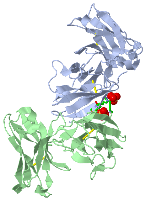
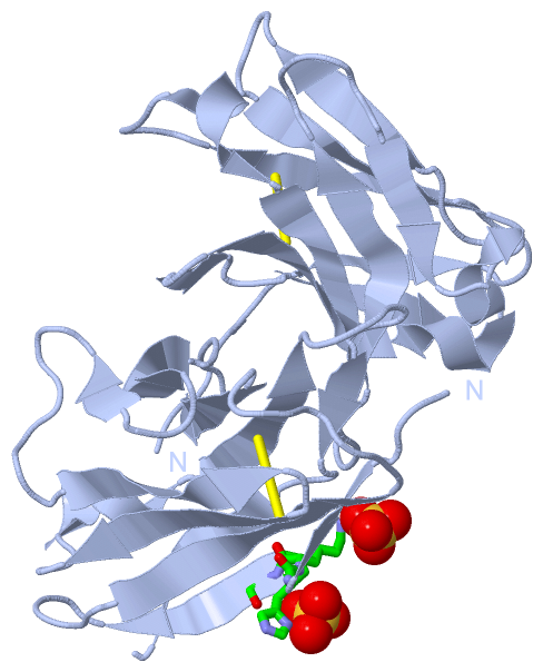
Chain A from PDB  Type:PROTEIN  Length:220
                                                                                                                                                                                                                                                            
               SCOP domains d1h8oa1 A:4-111 Immunoglobulin light chain kappa variable domain, VL-kappa                                  d1h8oa2 A:132-243 Immunoglobulin heavy chain variable domain, VH                                                 SCOP domains
               CATH domains 1h8oA01 A:4-132 Immunoglobulins                                                                              1h8oA02 A:133-235 Immunoglobulins                                                                      -------- CATH domains
               Pfam domains ---------------------------------------------------------------------------------------------------------------------------------------------------------------------------------------------------------------------------- Pfam domains
         Sec.struct. author ...eee...eeee.....eeeeeee.......eeeeee......eeeee...ee.......eeeeee..eeeeee...hhhhh.eeeeee......ee...eeeee..........eeee.....eeeeee....hhhhh.eeeeee.....eeeeeee......eee.hhhh....eeee....eeeeee...hhhhheeeeeee..ee...eeeee.. Sec.struct. author
                 SAPs(SNPs) ---------------------------------------------------------------------------------------------------------------------------------------------------------------------------------------------------------------------------- SAPs(SNPs)
                    PROSITE ---------------------------------------------------------------------------------------------------------------------------------------------------------------------------------------------------------------------------- PROSITE
                 Transcript ---------------------------------------------------------------------------------------------------------------------------------------------------------------------------------------------------------------------------- Transcript
                 1h8o A   4 DIVLTQSHKFMSTSVGDRVSITCKASQDVGTAVAWYQQKPGQSPKLLIYWASTRHTGVPDRFTGSGSGTDFTLTISNVQSEDLADYFCQQYSSYPLTFGAGTKLELKRQVQLQEPGGELVRPGASVKLSCKASGYTFTSYWINWVKQRPGQGLEWIGNIYPSDSYTNYNQKFKDKATLTVDKSSSTAYMQLSSLTSEDSAVYFCARWGYWGQGTLVTVSA 243
                                    13        23        33        43        53        63        73        83        93       103       133       143       153       163       173       183       193       203       213       223       233       243
                                                                                                                                     111|                                                                                                               
                                                                                                                                      132                                                                                                               

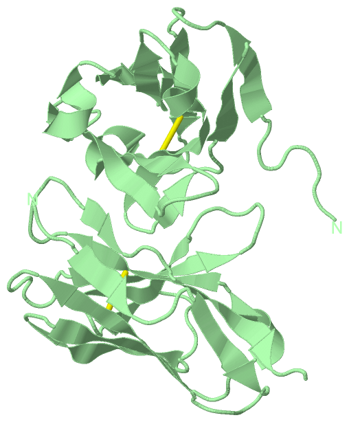
Chain B from PDB  Type:PROTEIN  Length:220
                                                                                                                                                                                                                                                            
               SCOP domains d1h8ob1 B:4-112 Immunoglobulin light chain kappa variable domain, VL-kappa                                   d1h8ob2 B:133-243 Immunoglobulin heavy chain variable domain, VH                                                SCOP domains
               CATH domains 1h8oB01 B:4-111 Immunoglobulins                                                                             -1h8oB02 B:133-231 Immunoglobulins                                                                  ------------ CATH domains
               Pfam domains ---------------------------------------------------------------------------------------------------------------------------------------------------------------------------------------------------------------------------- Pfam domains
         Sec.struct. author ...eee...eeee.....eeeeeee.......eeeeee......eeeee...ee.......eeeeee..eeeeee...hhhhh.eeeeee......ee...eeeee..........eeee.....eeeeee....hhhhh.eeeeee.....eeeeeee......eee.hhhh..eeeeeehhh.eeeeee...hhhhheeeeeee..ee...eeeee.. Sec.struct. author
                 SAPs(SNPs) ---------------------------------------------------------------------------------------------------------------------------------------------------------------------------------------------------------------------------- SAPs(SNPs)
                    PROSITE ---------------------------------------------------------------------------------------------------------------------------------------------------------------------------------------------------------------------------- PROSITE
                 Transcript ---------------------------------------------------------------------------------------------------------------------------------------------------------------------------------------------------------------------------- Transcript
                 1h8o B   4 DIVLTQSHKFMSTSVGDRVSITCKASQDVGTAVAWYQQKPGQSPKLLIYWASTRHTGVPDRFTGSGSGTDFTLTISNVQSEDLADYFCQQYSSYPLTFGAGTKLELKRGVQLQEPGGELVRPGASVKLSCKASGYTFTSYWINWVKQRPGQGLEWIGNIYPSDSYTNYNQKFKDKATLTVDKSSSTAYMQLSSLTSEDSAVYFCARWGYWGQGTLVTVSA 243
                                    13        23        33        43        53        63        73        83        93       103       133       143       153       163       173       183       193       203       213       223       233       243
                                                                                                                                      112|                                                                                                              
                                                                                                                                       133                                                                                                              

   Legend:   → Mismatch (orange background)
  - → Gap (green background, '-', border residues have a numbering label)
    → Modified Residue (blue background, lower-case, 'x' indicates undefined single-letter code, labelled with number + name)
  x → Chemical Group (purple background, 'x', labelled with number + name, e.g. ACE or NH2)
  extra numbering lines below/above indicate numbering irregularities and modified residue names etc., number ends below/above '|'

 Classification and Annotation

(-) SCOP Domains  (2, 4)

Asymmetric Unit

(-) CATH Domains  (1, 4)

Asymmetric Unit
(-)
Class: Mainly Beta (13760)

(-) Pfam Domains  (0, 0)

(no "Pfam Domain" information available for 1H8O)

(-) Gene Ontology  (0, 0)

Asymmetric Unit(hide GO term definitions)
    (no "Gene Ontology" information available for 1H8O)

 Visualization

(-) Interactive Views

Asymmetric Unit
  Complete Structure
    Jena3D(integrated viewing of ligand, site, SAP, PROSITE, SCOP information)
    WebMol | AstexViewer[tm]@PDBe
(Java Applets, require no local installation except for Java; loading may be slow)
    STRAP
(Java WebStart application, automatic local installation, requires Java; full application with system access!)
    RasMol
(require local installation)
    Molscript (VRML)
(requires installation of a VRML viewer; select preferred view via VRML and generate a mono or stereo PDF format file)
 
  Ligands, Modified Residues, Ions
    SO4  [ RasMol | Jena3D ]  +environment [ RasMol | Jena3D ]
 
  Sites
    AC1  [ RasMol ]  +environment [ RasMol ]
    AC2  [ RasMol ]  +environment [ RasMol ]
 
  Cis Peptide Bonds
    Glu A:137 - Pro A:138   [ RasMol ]  
    Glu B:137 - Pro B:138   [ RasMol ]  
    Tyr A:97 - Pro A:98   [ RasMol ]  
    Tyr B:97 - Pro B:98   [ RasMol ]  
 
Biological Units
  Complete Structure
    Biological Unit 1  [ Jena3D ]
    Biological Unit 2  [ Jena3D ]

(-) Still Images

Jmol
  protein: cartoon or spacefill or dots and stick; nucleic acid: cartoon and stick; ligands: spacefill; active site: stick
Molscript
  protein, nucleic acid: cartoon; ligands: spacefill; active site: ball and stick

 Databases and Analysis Tools

(-) Databases

Access by PDB/NDB ID
  1h8o
    Family and Domain InformationProDom | SYSTERS
    General Structural InformationGlycoscienceDB | MMDB | NDB | OCA | PDB | PDBe | PDBj | PDBsum | PDBWiki | PQS | PROTEOPEDIA
    Orientation in MembranesOPM
    Protein SurfaceSURFACE
    Secondary StructureDSSP (structure derived) | HSSP (homology derived)
    Structural GenomicsGeneCensus
    Structural NeighboursCE | VAST
    Structure ClassificationCATH | Dali | SCOP
    Validation and Original DataBMRB Data View | BMRB Restraints Grid | EDS | PROCHECK | RECOORD | WHAT_CHECK
 
Access by UniProt ID/Accession number
  (no 'UniProt ID/Accession number' available) |
    Comparative Protein Structure ModelsModBase
    Genomic InformationEnsembl
    Protein-protein InteractionDIP
    Sequence, Family and Domain InformationInterPro | Pfam | SMART | UniProt
 
Access by Enzyme Classificator   (EC Number)
  (no 'Enzyme Classificator' available)
    General Enzyme InformationBRENDA | EC-PDB | Enzyme | IntEnz
    PathwayKEGG | MetaCyc
 
Access by Disease Identifier   (MIM ID)
  (no 'MIM ID' available)
    Disease InformationOMIM
 
Access by GenAge ID
  (no 'GenAge ID' available)
    Age Related InformationGenAge

(-) Analysis Tools

Access by PDB/NDB ID
    Domain InformationXDom
    Interatomic Contacts of Structural UnitsCSU
    Ligand-protein ContactsLPC
    Protein CavitiescastP
    Sequence and Secondary StructurePDBCartoon
    Structure AlignmentSTRAP(Java WebStart application, automatic local installation, requires Java; full application with system access!)
    Structure and Sequence BrowserSTING
 
Access by UniProt ID/Accession number
  (no 'UniProt ID/Accession number' available) |
    Protein Disorder PredictionDisEMBL | FoldIndex | GLOBPLOT

 Related Entries

(-) Entries Sharing at Least One Protein Chain (UniProt ID)

(no "Entries Sharing at Least One Protein Chain" available for 1H8O)

(-) Related Entries Specified in the PDB File

1h8n THREE-DIMENSIONAL STRUCTURE OF ANTI-AMPICILLIN SINGLE CHAIN FV FRAGMENT FROM PHAGE-DISPLAYED MURINE ANTIBODY LIBRARIES
1h8s THREE-DIMENSIONAL STRUCTURE OF ANTI-AMPICILLIN SINGLE CHAIN FV FRAGMENT COMPLEXED WITH THE HAPTEN.