Show PDB file:   
         Plain Text   HTML   (compressed file size)
QuickSearch:   
by PDB,NDB,UniProt,PROSITE Code or Search Term(s)  
(-)Asym./Biol. Unit - manually
(-)Asym./Biol. Unit
(-)Asym./Biol. Unit - sites
collapse expand < >
Image Asym./Biol. Unit - manually
Asym./Biol. Unit - manually  (Jmol Viewer)
Image Asym./Biol. Unit
Asym./Biol. Unit  (Jmol Viewer)
Image Asym./Biol. Unit - sites
Asym./Biol. Unit - sites  (Jmol Viewer)

(-) Description

Title :  THREE-DIMENSIONAL STRUCTURE OF ANTI-AMPICILLIN SINGLE CHAIN FV FRAGMENT FROM PHAGE-DISPLAYED MURINE ANTIBODY LIBRARIES
 
Authors :  S. Jung, S. Spinelli, B. Schimmele, A. Honegger, L. Pugliese, C. Cambillau, A. Pluckthun
Date :  14 Feb 01  (Deposition) - 02 Aug 01  (Release) - 24 Feb 09  (Revision)
Method :  X-RAY DIFFRACTION
Resolution :  1.87
Chains :  Asym./Biol. Unit :  A
Keywords :  Antibody, Framework (Keyword Search: [Gene Ontology, PubMed, Web (Google))
 
Reference :  S. Jung, S. Spinelli, B. Schimmele, A. Honegger, L. Pugliese, C. Cambillau, A. Pluckthun
The Importance Of Framework Residues H6, H7 And H10 In Antibody Heavy Chains: Experimental Evidence For A New Structural Subclassification Of Antibody V(H) Domains
J. Mol. Biol. V. 309 701 2001
PubMed-ID: 11397090  |  Reference-DOI: 10.1006/JMBI.2001.4665

(-) Compounds

Molecule 1 - MUTANT AL2 6E7S9G
    ChainsA
    EngineeredYES
    Expression SystemESCHERICHIA COLI
    Expression System Taxid562
    FragmentFV FRAGMENT
    Organism ScientificMUS MUSCULUS
    Organism Taxid10090
    Other DetailsSINGLE CHAIN FV ANTIBODY IN WHICH THE VL AND VH FRAGMENTS ARE JOINED BY A GGGGSGGGGSGGGGSGGGGS LINKER

 Structural Features

(-) Chains, Units

  1
Asymmetric/Biological Unit A

Summary Information (see also Sequences/Alignments below)

(-) Ligands, Modified Residues, Ions  (2, 3)

Asymmetric/Biological Unit (2, 3)
No.NameCountTypeFull Name
1GOL2Ligand/IonGLYCEROL
2SO41Ligand/IonSULFATE ION

(-) Sites  (3, 3)

Asymmetric Unit (3, 3)
No.NameEvidenceResiduesDescription
1AC1SOFTWAREARG A:21 , SER A:23 , SER A:156 , GLY A:157 , HOH A:2103BINDING SITE FOR RESIDUE SO4 A1002
2AC2SOFTWARESER A:79 , TYR A:158 , THR A:159 , SER A:162 , TYR A:163BINDING SITE FOR RESIDUE GOL A1000
3AC3SOFTWARETYR A:191 , LYS A:196 , HOH A:2075 , HOH A:2079BINDING SITE FOR RESIDUE GOL A1001

(-) SS Bonds  (2, 2)

Asymmetric/Biological Unit
No.Residues
1A:26 -A:91
2A:153 -A:227

(-) Cis Peptide Bonds  (1, 1)

Asymmetric/Biological Unit
No.Residues
1Tyr A:97 -Pro A:98

 Sequence-Structure Mapping

(-) SAPs(SNPs)/Variants  (0, 0)

(no "SAP(SNP)/Variant" information available for 1H8N)

(-) PROSITE Motifs  (0, 0)

(no "PROSITE Motif" information available for 1H8N)

(-) Exons   (0, 0)

(no "Exon" information available for 1H8N)

(-) Sequences/Alignments

Asymmetric/Biological Unit
   Reformat: Number of residues per line =  ('0' or empty: single-line sequence representation)
  Number of residues per labelling interval =   
  UniProt sequence: complete  aligned part    
   Show mapping: SCOP domains CATH domains Pfam domains Secondary structure (by author)
SAPs(SNPs) PROSITE motifs Exons
(details for a mapped element are shown in a popup box when the mouse pointer rests over it)
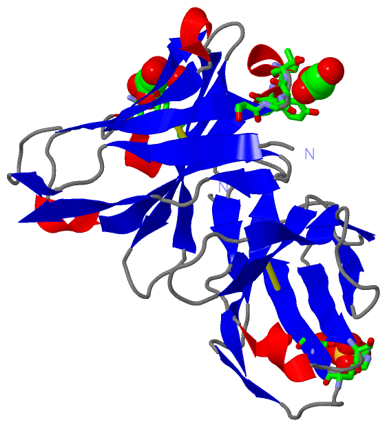
Chain A from PDB  Type:PROTEIN  Length:219
                                                                                                                                                                                                                                                           
               SCOP domains d1h8na1 A:3-109 Immunoglobulin light chain kappa variable domain, VL-kappa                                 d1h8na2 A:132-243 Immunoglobulin heavy chain variable domain, VH                                                 SCOP domains
               CATH domains -1h8nA01 A:4-134 Immunoglobulins                                                                              1h8nA02 A:135-233 Immunoglobulins                                                                  ---------- CATH domains
               Pfam domains --------------------------------------------------------------------------------------------------------------------------------------------------------------------------------------------------------------------------- Pfam domains
         Sec.struct. author ....ee....eee......eeeeeee.......eeeeee......eeeee...ee.......eeeeee..eeeeee...hhhhh.eeeeee......ee...eeee...eeeee...ee.....eeeeeeee..hhhhh.eeeeee.....eeeeeeee....eeee.hhhh..eeeeeehhh.eeeeee...hhhhheeeeeee..ee...eeeee.. Sec.struct. author
                 SAPs(SNPs) --------------------------------------------------------------------------------------------------------------------------------------------------------------------------------------------------------------------------- SAPs(SNPs)
                    PROSITE --------------------------------------------------------------------------------------------------------------------------------------------------------------------------------------------------------------------------- PROSITE
                 Transcript --------------------------------------------------------------------------------------------------------------------------------------------------------------------------------------------------------------------------- Transcript
                 1h8n A   3 KDIVLTQSHKFMSTSVGDRVSITCKASQDVGTAVAWYQQKPGQSPKLLIYWASTRHTGVPDRFTGSGSGTDFTLTISNVQSEDLADYFCQQYSSYPLTFGAGTKLELQVQLQESGGELVRPGASVKLSCKASGYTFTSYWINWVKQRPGQGLEWIGNIYPSDSYTNYNQKFKDKATLTVDKSSSTAYMQLSSLTSEDSAVYFCARWGYWGQGTLVTVSA 243
                                    12        22        32        42        52        62        72        82        92       102      |134       144       154       164       174       184       194       204       214       224       234         
                                                                                                                                    109|                                                                                                               
                                                                                                                                     132                                                                                                               

   Legend:   → Mismatch (orange background)
  - → Gap (green background, '-', border residues have a numbering label)
    → Modified Residue (blue background, lower-case, 'x' indicates undefined single-letter code, labelled with number + name)
  x → Chemical Group (purple background, 'x', labelled with number + name, e.g. ACE or NH2)
  extra numbering lines below/above indicate numbering irregularities and modified residue names etc., number ends below/above '|'

 Classification and Annotation

(-) SCOP Domains  (2, 2)

Asymmetric/Biological Unit

(-) CATH Domains  (1, 2)

Asymmetric/Biological Unit
(-)
Class: Mainly Beta (13760)

(-) Pfam Domains  (0, 0)

(no "Pfam Domain" information available for 1H8N)

(-) Gene Ontology  (0, 0)

Asymmetric/Biological Unit(hide GO term definitions)
    (no "Gene Ontology" information available for 1H8N)

 Visualization

(-) Interactive Views

Asymmetric/Biological Unit
  Complete Structure
    Jena3D(integrated viewing of ligand, site, SAP, PROSITE, SCOP information)
    WebMol | AstexViewer[tm]@PDBe
(Java Applets, require no local installation except for Java; loading may be slow)
    STRAP
(Java WebStart application, automatic local installation, requires Java; full application with system access!)
    RasMol
(require local installation)
    Molscript (VRML)
(requires installation of a VRML viewer; select preferred view via VRML and generate a mono or stereo PDF format file)
 
  Ligands, Modified Residues, Ions
    GOL  [ RasMol | Jena3D ]  +environment [ RasMol | Jena3D ]
    SO4  [ RasMol | Jena3D ]  +environment [ RasMol | Jena3D ]
 
  Sites
    AC1  [ RasMol ]  +environment [ RasMol ]
    AC2  [ RasMol ]  +environment [ RasMol ]
    AC3  [ RasMol ]  +environment [ RasMol ]
 
  Cis Peptide Bonds
    Tyr A:97 - Pro A:98   [ RasMol ]  
 

(-) Still Images

Jmol
  protein: cartoon or spacefill or dots and stick; nucleic acid: cartoon and stick; ligands: spacefill; active site: stick
Molscript
  protein, nucleic acid: cartoon; ligands: spacefill; active site: ball and stick

 Databases and Analysis Tools

(-) Databases

Access by PDB/NDB ID
  1h8n
    Family and Domain InformationProDom | SYSTERS
    General Structural InformationGlycoscienceDB | MMDB | NDB | OCA | PDB | PDBe | PDBj | PDBsum | PDBWiki | PQS | PROTEOPEDIA
    Orientation in MembranesOPM
    Protein SurfaceSURFACE
    Secondary StructureDSSP (structure derived) | HSSP (homology derived)
    Structural GenomicsGeneCensus
    Structural NeighboursCE | VAST
    Structure ClassificationCATH | Dali | SCOP
    Validation and Original DataBMRB Data View | BMRB Restraints Grid | EDS | PROCHECK | RECOORD | WHAT_CHECK
 
Access by UniProt ID/Accession number
  (no 'UniProt ID/Accession number' available) |
    Comparative Protein Structure ModelsModBase
    Genomic InformationEnsembl
    Protein-protein InteractionDIP
    Sequence, Family and Domain InformationInterPro | Pfam | SMART | UniProt
 
Access by Enzyme Classificator   (EC Number)
  (no 'Enzyme Classificator' available)
    General Enzyme InformationBRENDA | EC-PDB | Enzyme | IntEnz
    PathwayKEGG | MetaCyc
 
Access by Disease Identifier   (MIM ID)
  (no 'MIM ID' available)
    Disease InformationOMIM
 
Access by GenAge ID
  (no 'GenAge ID' available)
    Age Related InformationGenAge

(-) Analysis Tools

Access by PDB/NDB ID
    Domain InformationXDom
    Interatomic Contacts of Structural UnitsCSU
    Ligand-protein ContactsLPC
    Protein CavitiescastP
    Sequence and Secondary StructurePDBCartoon
    Structure AlignmentSTRAP(Java WebStart application, automatic local installation, requires Java; full application with system access!)
    Structure and Sequence BrowserSTING
 
Access by UniProt ID/Accession number
  (no 'UniProt ID/Accession number' available) |
    Protein Disorder PredictionDisEMBL | FoldIndex | GLOBPLOT

 Related Entries

(-) Entries Sharing at Least One Protein Chain (UniProt ID)

(no "Entries Sharing at Least One Protein Chain" available for 1H8N)

(-) Related Entries Specified in the PDB File

1h8o THREE-DIMENSIONAL STRUCTURE OF ANTI-AMPICILLIN SINGLE CHAIN FV FRAGMENT.
1h8s THREE-DIMENSIONAL STRUCTURE OF ANTI-AMPICILLIN SINGLE CHAIN FV FRAGMENT COMPLEXED WITH THE HAPTEN.