|
|
|
|
 Description
|
|
Compounds
|
||||||||||||||||||||||||||||||||||||||||||||||||||||||||||||||||||||||||||||||||||||||||||||||
Chains, Units
Summary Information (see also Sequences/Alignments below) |
Ligands, Modified Residues, Ions (2, 2)| Asymmetric Unit (2, 2) Biological Unit 1 (2, 4) |
Sites (2, 2)
Asymmetric Unit (2, 2)
|
SS Bonds (0, 0)| (no "SS Bond" information available for 1H2S) |
Cis Peptide Bonds (0, 0)| (no "Cis Peptide Bond" information available for 1H2S) |
SAPs(SNPs)/Variants (0, 0)| (no "SAP(SNP)/Variant" information available for 1H2S) |
PROSITE Motifs (2, 2)
Asymmetric Unit (2, 2)
|
||||||||||||||||||||||||||||||||||||||||||||||||||||||||||||||||
Exons (0, 0)| (no "Exon" information available for 1H2S) |
Sequences/Alignments
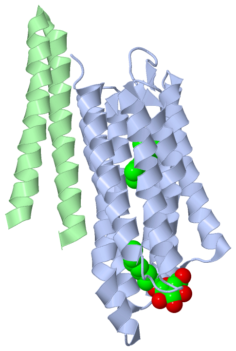
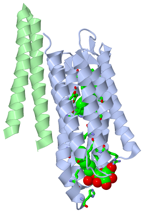
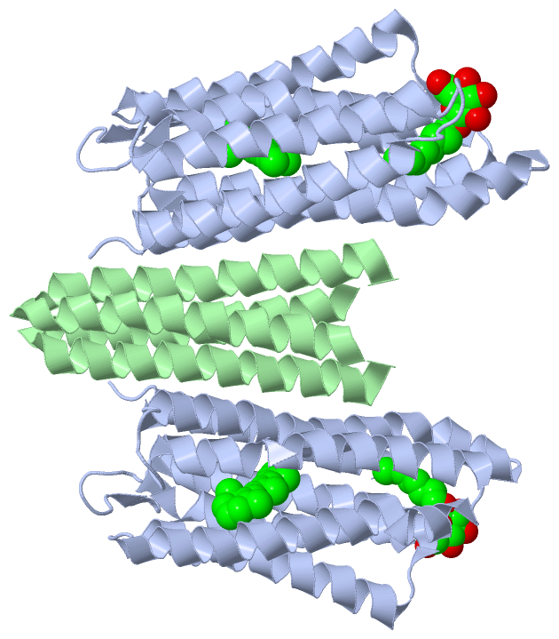
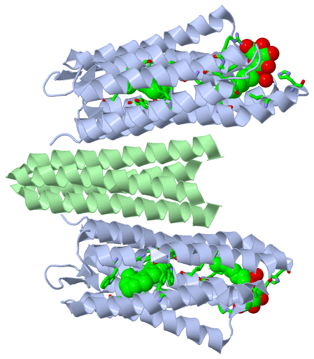
Asymmetric UnitChain A from PDB Type:PROTEIN Length:225 aligned with BACS2_NATPH | P42196 from UniProtKB/Swiss-Prot Length:239 Alignment length:225 10 20 30 40 50 60 70 80 90 100 110 120 130 140 150 160 170 180 190 200 210 220 BACS2_NATPH 1 MVGLTTLFWLGAIGMLVGTLAFAWAGRDAGSGERRYYVTLVGISGIAAVAYVVMALGVGWVPVAERTVFAPRYIDWILTTPLIVYFLGLLAGLDSREFGIVITLNTVVMLAGFAGAMVPGIERYALFGMGAVAFLGLVYYLVGPMTESASQRSSGIKSLYVRLRNLTVILWAIYPFIWLLGPPGVALLTPTVDVALIVYLDLVTKVGFGFIALDAAATLRAEHGE 225 SCOP domains d1h2sa_ A: Sensory rhodopsin II SCOP domains CATH domains 1h2sA00 A:1-225 Rhopdopsin 7-helix transmembrane proteins CATH domains Pfam domains --------------------------------------------------------------------------------------------------------------------------------------------------------------------------------------------------------------------------------- Pfam domains SAPs(SNPs) --------------------------------------------------------------------------------------------------------------------------------------------------------------------------------------------------------------------------------- SAPs(SNPs) PROSITE -----------------------------------------------------------------------BACTERIAL_OPS----------------------------------------------------------------------------------------------------------------BACTERIAL_OP----------------- PROSITE Transcript --------------------------------------------------------------------------------------------------------------------------------------------------------------------------------------------------------------------------------- Transcript 1h2s A 1 MVGLTTLFWLGAIGMLVGTLAFAWAGRDAGSGERRYYVTLVGISGIAAVAYVVMALGVGWVPVAERTVFAPRYIDWILTTPLIVYFLGLLAGLDSREFGIVITLNTVVMLAGFAGAMVPGIERYALFGMGAVAFLGLVYYLVGPMTESASQRSSGIKSLYVRLRNLTVILWAIYPFIWLLGPPGVALLTPTVDVALIVYLDLVTKVGFGFIALDAAATLRAEHGE 225 10 20 30 40 50 60 70 80 90 100 110 120 130 140 150 160 170 180 190 200 210 220 Chain B from PDB Type:PROTEIN Length:60 aligned with HTR2_NATPH | P42259 from UniProtKB/Swiss-Prot Length:534 Alignment length:60 32 42 52 62 72 82 HTR2_NATPH 23 GAVFIFVGALTVLFGAIAYGEVTAAAATGDAAAVQEAAVSAILGLIILLGINLGLVAATL 82 SCOP domains d1h2sb_ B: Sensory rhodopsin II transducer, Htr2 SCOP domains CATH domains 1h2sB00 B:23-82 Helix hairpin bin CATH domains Pfam domains ------------------------------------------------------------ Pfam domains SAPs(SNPs) ------------------------------------------------------------ SAPs(SNPs) PROSITE ------------------------------------------------------------ PROSITE Transcript ------------------------------------------------------------ Transcript 1h2s B 23 GAVFIFVGALTVLFGAIAYGEVTAAAATGDAAAVQEAAVSAILGLIILLGINLGLVAATL 82 32 42 52 62 72 82
|
||||||||||||||||||||
SCOP Domains (2, 2)
Asymmetric Unit
|
CATH Domains (2, 2)
Asymmetric Unit
 |
Pfam Domains (0, 0)| (no "Pfam Domain" information available for 1H2S) |
Gene Ontology (14, 20)|
Asymmetric Unit(hide GO term definitions) Chain A (BACS2_NATPH | P42196)
Chain B (HTR2_NATPH | P42259)
|
||||||||||||||||||||||||||||||||||||||||||||||||||||||||||||||||||||||||||||||||||||||||||||||||||||||||||||||||||||||||||||||||||||||||||||||||||||||||||||
Interactive Views
|
||||||||||||||||||||||||||||||||||||||||||||||||||||||||||||||||||||||||||||||||||||||||||||||||||||||||||||||||||||||||||||||||||||||||||||||||||||||
Still Images
|
||||||||||||||||
Databases
|
||||||||||||||||||||||||||||||||||||||||||||||||||||||||||||||||||||||||||||||||||||||||||||||||||||||||||||||||||||||||||||||||||||||||||||||||||||||||||||||||||||||||||||||||||||||||||
Analysis Tools
|
||||||||||||||||||||||||||||||||||||||||||||||||||||||||||||||||||||||||
Entries Sharing at Least One Protein Chain (UniProt ID)
Related Entries Specified in the PDB File
|
|