Show PDB file:   
         Plain Text   HTML   (compressed file size)
QuickSearch:   
by PDB,NDB,UniProt,PROSITE Code or Search Term(s)  
(-)Asym./Biol. Unit - manually
(-)Asym./Biol. Unit
(-)Asym./Biol. Unit - sites
collapse expand < >
Image Asym./Biol. Unit - manually
Asym./Biol. Unit - manually  (Jmol Viewer)
Image Asym./Biol. Unit
Asym./Biol. Unit  (Jmol Viewer)
Image Asym./Biol. Unit - sites
Asym./Biol. Unit - sites  (Jmol Viewer)

(-) Description

Title :  HIGH RESOLUTION CRYSTAL STRUCTURE OF THE FV FRAGMENT FROM AN ANTI-DANSYL SWITCH VARIANT ANTIBODY IGG2A(S) CRYSTALLIZED AT PH 5.25
 
Authors :  M. Nakasako, H. Takahashi, I. Shimada, Y. Arata
Date :  14 Jul 98  (Deposition) - 26 Jul 99  (Release) - 24 Feb 09  (Revision)
Method :  X-RAY DIFFRACTION
Resolution :  1.45
Chains :  Asym./Biol. Unit :  H,L
Keywords :  Fv Fragment, Immunoglobulin (Keyword Search: [Gene Ontology, PubMed, Web (Google))
 
Reference :  M. Nakasako, H. Takahashi, N. Shimba, I. Shimada, Y. Arata
The Ph-Dependent Structural Variation Of Complementarity-Determining Region H3 In The Crystal Structures Of The Fv Fragment From An Anti-Dansyl Monoclonal Antibody.
J. Mol. Biol. V. 291 117 1999
PubMed-ID: 10438610  |  Reference-DOI: 10.1006/JMBI.1999.2931
(for further references see the PDB file header)

(-) Compounds

Molecule 1 - ANTI-DANSYL IMMUNOGLOBULIN IGG2A(S)
    Cell LineHYBRIDOMA
    ChainsL
    FragmentFV FRAGMENT (VH AND VL DOMAINS)
    Organism CommonHOUSE MOUSE
    Organism ScientificMUS MUSCULUS
    Organism Taxid10090
    SynonymANTI-DANSYL FV FRAGMENT
 
Molecule 2 - ANTI-DANSYL IMMUNOGLOBULIN IGG2A(S)
    Cell LineHYBRIDOMA
    ChainsH
    FragmentFV FRAGMENT (VH AND VL DOMAINS)
    Organism CommonHOUSE MOUSE
    Organism ScientificMUS MUSCULUS
    Organism Taxid10090
    SynonymANTI-DANSYL FV FRAGMENT

 Structural Features

(-) Chains, Units

  12
Asymmetric/Biological Unit HL

Summary Information (see also Sequences/Alignments below)

(-) Ligands, Modified Residues, Ions  (1, 4)

Asymmetric/Biological Unit (1, 4)
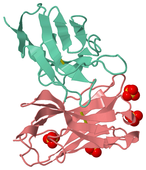
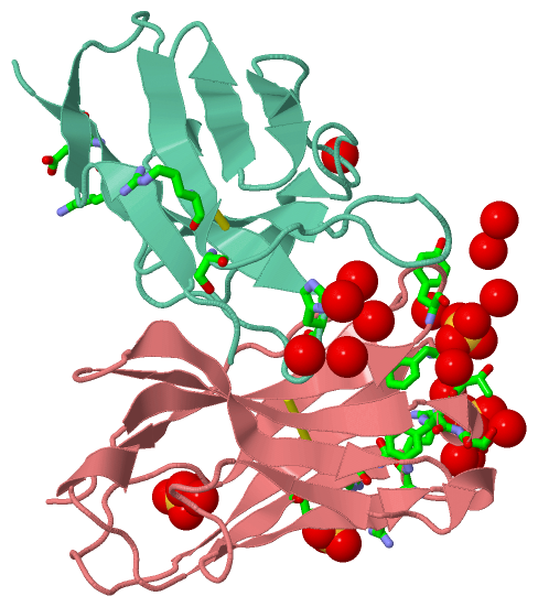
No.NameCountTypeFull Name
1SO44Ligand/IonSULFATE ION

(-) Sites  (4, 4)

Asymmetric Unit (4, 4)
No.NameEvidenceResiduesDescription
1AC1SOFTWAREPHE H:27 , THR H:28 , TYR H:97 , HOH H:570 , HOH H:618 , HOH H:652 , HOH H:682 , ARG L:24BINDING SITE FOR RESIDUE SO4 H 500
2AC2SOFTWARELYS H:75 , ARG H:77 , TYR H:79 , HOH H:608 , HOH H:614 , SER L:26BINDING SITE FOR RESIDUE SO4 H 501
3AC3SOFTWAREHOH H:694 , HIS L:93 , LYS L:103 , GLU L:105 , HOH L:117 , HOH L:128 , HOH L:139 , HOH L:214 , HOH L:256BINDING SITE FOR RESIDUE SO4 H 502
4AC4SOFTWAREPHE H:27 , PHE H:29 , SER H:30 , ARG H:76 , HOH H:599 , HOH H:609 , HOH H:632 , HOH H:663 , HOH L:283BINDING SITE FOR RESIDUE SO4 H 503

(-) SS Bonds  (2, 2)

Asymmetric/Biological Unit
No.Residues
1H:22 -H:92
2L:23 -L:88

(-) Cis Peptide Bonds  (2, 2)

Asymmetric/Biological Unit
No.Residues
1Thr L:7 -Pro L:8
2Val L:94 -Pro L:95

 Sequence-Structure Mapping

(-) SAPs(SNPs)/Variants  (0, 0)

(no "SAP(SNP)/Variant" information available for 1DLF)

(-) PROSITE Motifs  (0, 0)

(no "PROSITE Motif" information available for 1DLF)

(-) Exons   (0, 0)

(no "Exon" information available for 1DLF)

(-) Sequences/Alignments

Asymmetric/Biological Unit
   Reformat: Number of residues per line =  ('0' or empty: single-line sequence representation)
  Number of residues per labelling interval =   
  UniProt sequence: complete  aligned part    
   Show mapping: SCOP domains CATH domains Pfam domains Secondary structure (by author)
SAPs(SNPs) PROSITE motifs Exons
(details for a mapped element are shown in a popup box when the mouse pointer rests over it)
Chain H from PDB  Type:PROTEIN  Length:120
                                                                                                                                                        
               SCOP domains d1dlfh_ H: Immunoglobulin heavy chain variable domain, VH                                                                SCOP domains
               CATH domains 1dlfH00 H:1-114 Immunoglobulins                                                                                          CATH domains
               Pfam domains ------------------------------------------------------------------------------------------------------------------------ Pfam domains
         Sec.struct. author ..eeeee..........eeeeeeee...hhh.eeeeeeee...eeeeeeee..hhh...eee.......eeeeee....eeeeee....hhh.eeeeeeee.............eee... Sec.struct. author
                 SAPs(SNPs) ------------------------------------------------------------------------------------------------------------------------ SAPs(SNPs)
                    PROSITE ------------------------------------------------------------------------------------------------------------------------ PROSITE
                 Transcript ------------------------------------------------------------------------------------------------------------------------ Transcript
                 1dlf H   1 EVKLEESGGGLVQPGGSMKLSCATSGFTFSDAWMDWVRQSPEKGLEWVAEIRNKANNHATYYAESVKGRFTISRDDSKRRVYLQMNTLRAEDTGIYYCTGIYYHYPWFAYWGQGTLVTVS 114
                                    10        20        30        40        50  |||   57        67        77     |||84        94       104       114
                                                                              52A||                            82A||                                
                                                                               52B|                             82B|                                
                                                                                52C                              82C                                

Chain L from PDB  Type:PROTEIN  Length:113
 aligned with KV2A7_MOUSE | P01631 from UniProtKB/Swiss-Prot  Length:113

    Alignment length:113
                                    10        20        30        40        50        60        70        80        90       100       110   
          KV2A7_MOUSE     1 DVVMTQTPLSLPVSLGDQASISCRSSQSLVHSNGNTYLNWYLQKAGQSPKLLIYKVSNRFSGVPDRFSGSGSGTDFTLKISRVEAEDLGIYFCSQTTHVPPTFGGGTKLEIKR 113
               SCOP domains d1dlfl_ L: Immunoglobulin light chain kappa variable domain, VL-kappa                                             SCOP domains
               CATH domains 1dlfL00 L:1-108 Immunoglobulins                                                                                   CATH domains
               Pfam domains ----------------------------------------------------------------------------------------------------------------- Pfam domains
         Sec.struct. author ...eeee..eeee.....eeeeeee............eeeeee......eeee.............eeeeee..eeeeee....hhh.eeeeeee...........eeeee.. Sec.struct. author
                 SAPs(SNPs) ----------------------------------------------------------------------------------------------------------------- SAPs(SNPs)
                    PROSITE ----------------------------------------------------------------------------------------------------------------- PROSITE
                 Transcript ----------------------------------------------------------------------------------------------------------------- Transcript
                 1dlf L   1 DVVMTQTPLSLPVSLGNQASISCRSSQSLVHSNGNTYLHWYLQKPGQSPKLLIYKVSNRFSGVPDRFSGSGSGTDFTLKISRVEAEDLGVYFCSQSTHVPFTFGSGTKLEIKR 108
                                    10        20       27C||      35        45        55        65        75        85        95       105   
                                                     27A||||                                                                                 
                                                      27B|||                                                                                 
                                                       27C||                                                                                 
                                                        27D|                                                                                 
                                                         27E                                                                                 

   Legend:   → Mismatch (orange background)
  - → Gap (green background, '-', border residues have a numbering label)
    → Modified Residue (blue background, lower-case, 'x' indicates undefined single-letter code, labelled with number + name)
  x → Chemical Group (purple background, 'x', labelled with number + name, e.g. ACE or NH2)
  extra numbering lines below/above indicate numbering irregularities and modified residue names etc., number ends below/above '|'

 Classification and Annotation

(-) SCOP Domains  (2, 2)

Asymmetric/Biological Unit

(-) CATH Domains  (1, 2)

Asymmetric/Biological Unit
(-)
Class: Mainly Beta (13760)

(-) Pfam Domains  (0, 0)

(no "Pfam Domain" information available for 1DLF)

(-) Gene Ontology  (1, 1)

Asymmetric/Biological Unit(hide GO term definitions)
Chain L   (KV2A7_MOUSE | P01631)
molecular function
    GO:0003823    antigen binding    Interacting selectively and non-covalently with an antigen, any substance which is capable of inducing a specific immune response and of reacting with the products of that response, the specific antibody or specifically sensitized T-lymphocytes, or both. Binding may counteract the biological activity of the antigen.

 Visualization

(-) Interactive Views

Asymmetric/Biological Unit
  Complete Structure
    Jena3D(integrated viewing of ligand, site, SAP, PROSITE, SCOP information)
    WebMol | AstexViewer[tm]@PDBe
(Java Applets, require no local installation except for Java; loading may be slow)
    STRAP
(Java WebStart application, automatic local installation, requires Java; full application with system access!)
    RasMol
(require local installation)
    Molscript (VRML)
(requires installation of a VRML viewer; select preferred view via VRML and generate a mono or stereo PDF format file)
 
  Ligands, Modified Residues, Ions
    SO4  [ RasMol | Jena3D ]  +environment [ RasMol | Jena3D ]
 
  Sites
    AC1  [ RasMol ]  +environment [ RasMol ]
    AC2  [ RasMol ]  +environment [ RasMol ]
    AC3  [ RasMol ]  +environment [ RasMol ]
    AC4  [ RasMol ]  +environment [ RasMol ]
 
  Cis Peptide Bonds
    Thr L:7 - Pro L:8   [ RasMol ]  
    Val L:94 - Pro L:95   [ RasMol ]  
 

(-) Still Images

Jmol
  protein: cartoon or spacefill or dots and stick; nucleic acid: cartoon and stick; ligands: spacefill; active site: stick
Molscript
  protein, nucleic acid: cartoon; ligands: spacefill; active site: ball and stick

 Databases and Analysis Tools

(-) Databases

Access by PDB/NDB ID
  1dlf
    Family and Domain InformationProDom | SYSTERS
    General Structural InformationGlycoscienceDB | MMDB | NDB | OCA | PDB | PDBe | PDBj | PDBsum | PDBWiki | PQS | PROTEOPEDIA
    Orientation in MembranesOPM
    Protein SurfaceSURFACE
    Secondary StructureDSSP (structure derived) | HSSP (homology derived)
    Structural GenomicsGeneCensus
    Structural NeighboursCE | VAST
    Structure ClassificationCATH | Dali | SCOP
    Validation and Original DataBMRB Data View | BMRB Restraints Grid | EDS | PROCHECK | RECOORD | WHAT_CHECK
 
Access by UniProt ID/Accession number
  KV2A7_MOUSE | P01631
    Comparative Protein Structure ModelsModBase
    Genomic InformationEnsembl
    Protein-protein InteractionDIP
    Sequence, Family and Domain InformationInterPro | Pfam | SMART | UniProtKB/SwissProt
 
Access by Enzyme Classificator   (EC Number)
  (no 'Enzyme Classificator' available)
    General Enzyme InformationBRENDA | EC-PDB | Enzyme | IntEnz
    PathwayKEGG | MetaCyc
 
Access by Disease Identifier   (MIM ID)
  (no 'MIM ID' available)
    Disease InformationOMIM
 
Access by GenAge ID
  (no 'GenAge ID' available)
    Age Related InformationGenAge

(-) Analysis Tools

Access by PDB/NDB ID
    Domain InformationXDom
    Interatomic Contacts of Structural UnitsCSU
    Ligand-protein ContactsLPC
    Protein CavitiescastP
    Sequence and Secondary StructurePDBCartoon
    Structure AlignmentSTRAP(Java WebStart application, automatic local installation, requires Java; full application with system access!)
    Structure and Sequence BrowserSTING
 
Access by UniProt ID/Accession number
  KV2A7_MOUSE | P01631
    Protein Disorder PredictionDisEMBL | FoldIndex | GLOBPLOT (for more information see DisProt)

 Related Entries

(-) Entries Sharing at Least One Protein Chain (UniProt ID)

UniProtKB/Swiss-Prot
        KV2A7_MOUSE | P016311bfv 1cfv 1maj 1mak 1n4x 1svz 1wz1 2bfv 2dlf 2hkf 2hkh 2ipt 2iqa 2o5x 2o5y 2o5z 2r0w 2vq1

(-) Related Entries Specified in the PDB File

(no "Related Entries Specified in the PDB File" available for 1DLF)