| molecular function |
|---|
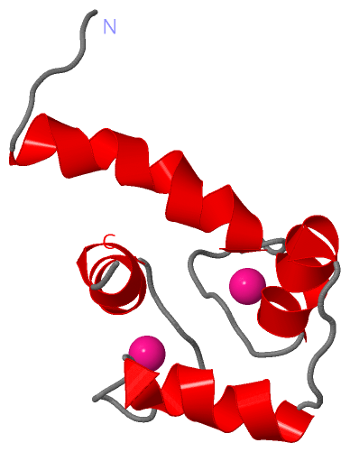
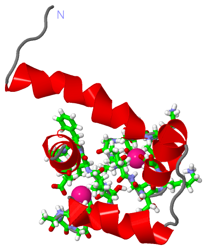
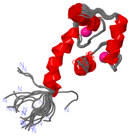
| | GO:0005509 | | calcium ion binding | | Interacting selectively and non-covalently with calcium ions (Ca2+). |
| | GO:0046872 | | metal ion binding | | Interacting selectively and non-covalently with any metal ion. |
| | GO:0005515 | | protein binding | | Interacting selectively and non-covalently with any protein or protein complex (a complex of two or more proteins that may include other nonprotein molecules). |
| biological process |
|---|
| | GO:0038095 | | Fc-epsilon receptor signaling pathway | | A series of molecular signals initiated by the binding of the Fc portion of immunoglobulin E (IgE) to an Fc-epsilon receptor on the surface of a signal-receiving cell, and ending with regulation of a downstream cellular process, e.g. transcription. The Fc portion of an immunoglobulin is its C-terminal constant region. |
| | GO:0000086 | | G2/M transition of mitotic cell cycle | | The mitotic cell cycle transition by which a cell in G2 commits to M phase. The process begins when the kinase activity of M cyclin/CDK complex reaches a threshold high enough for the cell cycle to proceed. This is accomplished by activating a positive feedback loop that results in the accumulation of unphosphorylated and active M cyclin/CDK complex. |
| | GO:0006936 | | muscle contraction | | A process in which force is generated within muscle tissue, resulting in a change in muscle geometry. Force generation involves a chemo-mechanical energy conversion step that is carried out by the actin/myosin complex activity, which generates force through ATP hydrolysis. |
| | GO:0060315 | | negative regulation of ryanodine-sensitive calcium-release channel activity | | Any process that decreases the activity of a ryanodine-sensitive calcium-release channel. The ryanodine-sensitive calcium-release channel catalyzes the transmembrane transfer of a calcium ion by a channel that opens when a ryanodine class ligand has been bound by the channel complex or one of its constituent parts. |
| | GO:0043388 | | positive regulation of DNA binding | | Any process that increases the frequency, rate or extent of DNA binding. DNA binding is any process in which a gene product interacts selectively with DNA (deoxyribonucleic acid). |
| | GO:0060316 | | positive regulation of ryanodine-sensitive calcium-release channel activity | | Any process that increases the activity of a ryanodine-sensitive calcium-release channel. The ryanodine-sensitive calcium-release channel catalyzes the transmembrane transfer of a calcium ion by a channel that opens when a ryanodine class ligand has been bound by the channel complex or one of its constituent parts. |
| | GO:0010880 | | regulation of release of sequestered calcium ion into cytosol by sarcoplasmic reticulum | | Any process that modulates the rate, frequency or extent of release of sequestered calcium ion into cytosol by the sarcoplasmic reticulum, the process in which the release of sequestered calcium ion by sarcoplasmic reticulum into cytosol occurs via calcium release channels. |
| | GO:0022400 | | regulation of rhodopsin mediated signaling pathway | | Any process that modulates the frequency, rate or extent of rhodopsin-mediated signaling. |
| cellular component |
|---|
| | GO:0005737 | | cytoplasm | | All of the contents of a cell excluding the plasma membrane and nucleus, but including other subcellular structures. |
| | GO:0005856 | | cytoskeleton | | Any of the various filamentous elements that form the internal framework of cells, and typically remain after treatment of the cells with mild detergent to remove membrane constituents and soluble components of the cytoplasm. The term embraces intermediate filaments, microfilaments, microtubules, the microtrabecular lattice, and other structures characterized by a polymeric filamentous nature and long-range order within the cell. The various elements of the cytoskeleton not only serve in the maintenance of cellular shape but also have roles in other cellular functions, including cellular movement, cell division, endocytosis, and movement of organelles. |
| | GO:0005829 | | cytosol | | The part of the cytoplasm that does not contain organelles but which does contain other particulate matter, such as protein complexes. |
| | GO:0005819 | | spindle | | The array of microtubules and associated molecules that forms between opposite poles of a eukaryotic cell during mitosis or meiosis and serves to move the duplicated chromosomes apart. |
| | GO:0000922 | | spindle pole | | Either of the ends of a spindle, where spindle microtubules are organized; usually contains a microtubule organizing center and accessory molecules, spindle microtubules and astral microtubules. |
| | GO:0008076 | | voltage-gated potassium channel complex | | A protein complex that forms a transmembrane channel through which potassium ions may cross a cell membrane in response to changes in membrane potential. |
 Description