|
|
|
|
 Description
|
|
Compounds
|
||||||||||||||||||||||||||||||||||||||||||||||||
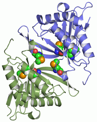
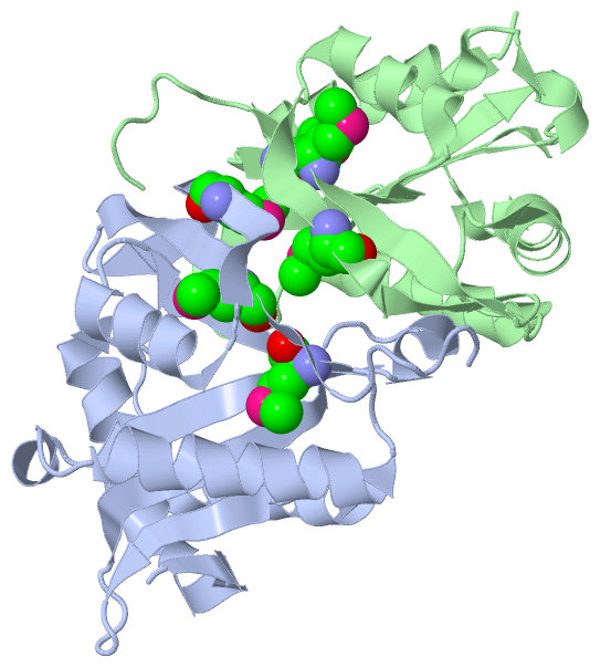
Chains, Units
Summary Information (see also Sequences/Alignments below) |
Ligands, Modified Residues, Ions (1, 6)
Asymmetric/Biological Unit (1, 6)
|
Sites (0, 0)| (no "Site" information available for 1YVO) |
SS Bonds (0, 0)| (no "SS Bond" information available for 1YVO) |
Cis Peptide Bonds (0, 0)| (no "Cis Peptide Bond" information available for 1YVO) |
SAPs(SNPs)/Variants (0, 0)| (no "SAP(SNP)/Variant" information available for 1YVO) |
PROSITE Motifs (1, 1)
Asymmetric/Biological Unit (1, 1)
|
||||||||||||||||||||||||
Exons (0, 0)| (no "Exon" information available for 1YVO) |
Sequences/Alignments
Asymmetric/Biological UnitChain A from PDB Type:PROTEIN Length:169 aligned with MDDA_PSEAE | Q9HUU7 from UniProtKB/Swiss-Prot Length:172 Alignment length:169 13 23 33 43 53 63 73 83 93 103 113 123 133 143 153 163 MDDA_PSEAE 4 SIRDAGVADLPGILAIYNDAVGNTTAIWNETPVDLANRQAWFDTRARQGYPILVASDAAGEVLGYASYGDWRPFEGFRGTVEHSVYVRDDQRGKGLGVQLLQALIERARAQGLHVMVAAIESGNAASIGLHRRLGFEISGQMPQVGQKFGRWLDLTFMQLNLDPTRSAP 172 SCOP domains d1yvoa1 A:4-172 Hypothetical protein PA4866 SCOP domains CATH domains ------------------------------------------------------------------------------------------------------------------------------------------------------------------------- CATH domains Pfam domains ------------------------------------------------------------------------------------------------------------------------------------------------------------------------- Pfam domains SAPs(SNPs) ------------------------------------------------------------------------------------------------------------------------------------------------------------------------- SAPs(SNPs) PROSITE GNAT PDB: - UniProt: 3-166 ------ PROSITE Transcript ------------------------------------------------------------------------------------------------------------------------------------------------------------------------- Transcript 1yvo A 4 SIRDAGVADLPGILAIYNDAVGNTTAIWNETPVDLANRQAWFDTRARQGYPILVASDAAGEVLGYASYGDWRPFEGFRGTVEHSVYVRDDQRGKGLGVQLLQALIERARAQGLHVmVAAIESGNAASIGLHRRLGFEISGQmPQVGQKFGRWLDLTFmQLNLDPTRSAP 172 13 23 33 43 53 63 73 83 93 103 113 | 123 133 143 | 153 163 119-MSE 145-MSE 161-MSE Chain B from PDB Type:PROTEIN Length:171 aligned with MDDA_PSEAE | Q9HUU7 from UniProtKB/Swiss-Prot Length:172 Alignment length:171 11 21 31 41 51 61 71 81 91 101 111 121 131 141 151 161 171 MDDA_PSEAE 2 SASIRDAGVADLPGILAIYNDAVGNTTAIWNETPVDLANRQAWFDTRARQGYPILVASDAAGEVLGYASYGDWRPFEGFRGTVEHSVYVRDDQRGKGLGVQLLQALIERARAQGLHVMVAAIESGNAASIGLHRRLGFEISGQMPQVGQKFGRWLDLTFMQLNLDPTRSAP 172 SCOP domains d1yvob_ B: automated matches SCOP domains CATH domains --------------------------------------------------------------------------------------------------------------------------------------------------------------------------- CATH domains Pfam domains --------------------------------------------------------------------------------------------------------------------------------------------------------------------------- Pfam domains SAPs(SNPs) --------------------------------------------------------------------------------------------------------------------------------------------------------------------------- SAPs(SNPs) PROSITE -GNAT PDB: B:3-166 UniProt: 3-166 ------ PROSITE Transcript --------------------------------------------------------------------------------------------------------------------------------------------------------------------------- Transcript 1yvo B 2 SASIRDAGVADLPGILAIYNDAVGNTTAIWNETPVDLANRQAWFDTRARQGYPILVASDAAGEVLGYASYGDWRPFEGFRGTVEHSVYVRDDQRGKGLGVQLLQALIERARAQGLHVmVAAIESGNAASIGLHRRLGFEISGQmPQVGQKFGRWLDLTFmQLNLDPTRSAP 172 11 21 31 41 51 61 71 81 91 101 111 121 131 141 | 151 161 171 119-MSE 145-MSE 161-MSE
|
||||||||||||||||||||
SCOP Domains (2, 2)
Asymmetric/Biological Unit
 |
CATH Domains (0, 0)| (no "CATH Domain" information available for 1YVO) |
Pfam Domains (0, 0)| (no "Pfam Domain" information available for 1YVO) |
Gene Ontology (7, 7)|
Asymmetric/Biological Unit(hide GO term definitions) Chain A,B (MDDA_PSEAE | Q9HUU7)
|
||||||||||||||||||||||||||||||||||||||||||||||||||||||||||||
Interactive Views
|
|||||||||||||||||||||||||||||||||||||||||||||||||||||||||||||||||||||||||||||||||||||||||||||||||||||||||||||||||||||
Still Images
|
||||||||||||||||
Databases
|
||||||||||||||||||||||||||||||||||||||||||||||||||||||||||||||||||||||||||||||||||||||||||||||||||||||||||||||||||||||||||||||||||||||||||||||||||||||||||||||||
Analysis Tools
|
|||||||||||||||||||||||||||||||||||||||||||||||||||||||||||||
Entries Sharing at Least One Protein Chain (UniProt ID)
Related Entries Specified in the PDB File
|
|