|
|
|
|
 Description
|
|
Compounds
|
||||||||||||||||||||||||||||||||||||||||||||||||||||||||
Chains, Units
Summary Information (see also Sequences/Alignments below) |
Ligands, Modified Residues, Ions (2, 2)| NMR Structure (2, 2) |
Sites (2, 2)
NMR Structure (2, 2)
|
SS Bonds (0, 0)| (no "SS Bond" information available for 1XXE) |
Cis Peptide Bonds (0, 0)| (no "Cis Peptide Bond" information available for 1XXE) |
SAPs(SNPs)/Variants (0, 0)| (no "SAP(SNP)/Variant" information available for 1XXE) |
PROSITE Motifs (0, 0)| (no "PROSITE Motif" information available for 1XXE) |
Exons (0, 0)| (no "Exon" information available for 1XXE) |
Sequences/Alignments
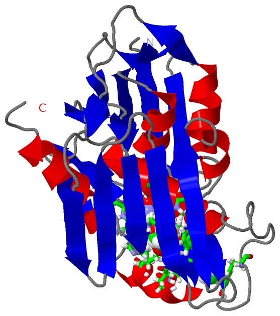
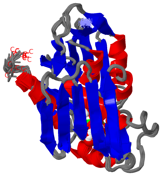
NMR StructureChain A from PDB Type:PROTEIN Length:268 aligned with LPXC_AQUAE | O67648 from UniProtKB/Swiss-Prot Length:282 Alignment length:268 12 22 32 42 52 62 72 82 92 102 112 122 132 142 152 162 172 182 192 202 212 222 232 242 252 262 LPXC_AQUAE 3 LEKTVKEKLSFEGVGIHTGEYSKLIIHPEKEGTGIRFFKNGVYIPARHEFVVHTNHSTDLGFKGQRIKTVEHILSVLHLLEITNVTIEVIGNEIPILDGSGWEFYEAIRKNILNQNREIDYFVVEEPIIVEDEGRLIKAEPSDTLEVTYEGEFKNFLGRQKFTFVEGNEEEIVLARTFCFDWEIEHIKKVGLGKGGSLKNTLVLGKDKVYNPEGLRYENEPVRHKVFDLIGDLYLLGSPVKGKFYSFRGGHSLNVKLVKELAKKQKLT 270 SCOP domains d1xxea1 A:3-127 UDP-3-O-[3-hydroxymyristoyl] N-acetylglucosamine deacetylase LpxC d1xxea2 A:128-270 UDP-3-O-[3-hydroxymyristoyl] N-acetylglucosamine deacetylase LpxC SCOP domains CATH domains 1xxeA01 A:3-122 lpxc deacetylase, domain 1 1xxeA02 A:123-270 lpxc deacetylase, domain 2 CATH domains Pfam domains LpxC-1xxeA01 A:3-268 -- Pfam domains SAPs(SNPs) ---------------------------------------------------------------------------------------------------------------------------------------------------------------------------------------------------------------------------------------------------------------------------- SAPs(SNPs) PROSITE ---------------------------------------------------------------------------------------------------------------------------------------------------------------------------------------------------------------------------------------------------------------------------- PROSITE Transcript ---------------------------------------------------------------------------------------------------------------------------------------------------------------------------------------------------------------------------------------------------------------------------- Transcript 1xxe A 3 LEKTVKEKLSFEGVGIHTGEYSKLIIHPEKEGTGIRFFKNGVYIPARHEFVVHTNHSTDLGFKGQRIKTVEHILSVLHLLEITNVTIEVIGNEIPILDGSGWEFYEAIRKNILNQNREIDYFVVEEPIIVEDEGRLIKAEPSDTLEVTYEGEFKNFLGRQKFTFVEGNEEEIVLARTFCFDWEIEHIKKVGLGKGGSLKNTLVLGKDKVYNPEGLRYENEPVRHKVFDLIGDLYLLGSPVKGKFYSFRGGHSLNVKLVKELAKKQKLT 270 12 22 32 42 52 62 72 82 92 102 112 122 132 142 152 162 172 182 192 202 212 222 232 242 252 262
|
||||||||||||||||||||
SCOP Domains (1, 2)
NMR Structure
 |
CATH Domains (2, 2)
NMR Structure
|
Pfam Domains (1, 1)
NMR Structure
|
Gene Ontology (6, 6)|
NMR Structure(hide GO term definitions) Chain A (LPXC_AQUAE | O67648)
|
||||||||||||||||||||||||||||||||||||||||||||||||
Interactive Views
|
||||||||||||||||||||||||||||||||||||||||||||||||||||||||||||||||||||||||||||||||||||||||||||||||||||||||||||||||||||||||||||||||||||
Still Images
|
||||||||||||||||
Databases
|
||||||||||||||||||||||||||||||||||||||||||||||||||||||||||||||||||||||||||||||||||||||||||||||||||||||||||||||||||||||||||||||||||||||||||||||||||||||||||||||||
Analysis Tools
|
|||||||||||||||||||||||||||||||||||||||||||||||||||||||||||||
Entries Sharing at Least One Protein Chain (UniProt ID)
Related Entries Specified in the PDB File
|
|