|
|
|
|
 Description
|
|
Compounds
|
||||||||||||||||||||||||||||||||||||||||||||||||||||
Chains, Units
Summary Information (see also Sequences/Alignments below) |
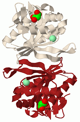
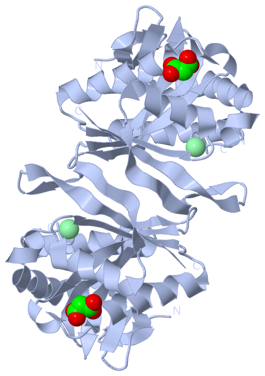
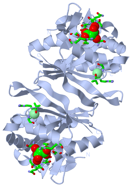
Ligands, Modified Residues, Ions (2, 2)| Asymmetric Unit (2, 2) Biological Unit 1 (1, 2) |
Sites (2, 2)
Asymmetric Unit (2, 2)
|
SS Bonds (0, 0)| (no "SS Bond" information available for 1S7F) |
Cis Peptide Bonds (0, 0)| (no "Cis Peptide Bond" information available for 1S7F) |
SAPs(SNPs)/Variants (0, 0)| (no "SAP(SNP)/Variant" information available for 1S7F) |
PROSITE Motifs (0, 0)| (no "PROSITE Motif" information available for 1S7F) |
Exons (0, 0)| (no "Exon" information available for 1S7F) |
Sequences/Alignments
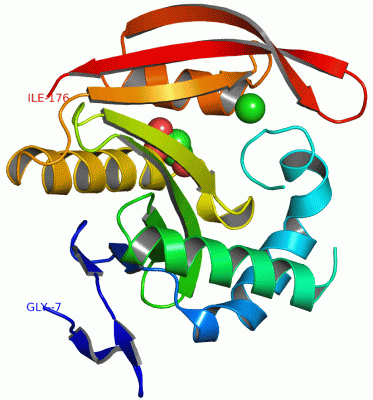
Asymmetric UnitChain A from PDB Type:PROTEIN Length:181 aligned with Q8ZPC0_SALTY | Q8ZPC0 from UniProtKB/TrEMBL Length:179 Alignment length:184 1 |2 12 22 32 42 52 62 72 82 92 102 112 122 132 142 152 162 172 Q8ZPC0_SALTY - --------MVEIIPVSTTLELRAADESHVPALHQLVLKNKAWLQQSLDWPQYVTSQEETRKHVQGNILLHQRGYAKMYLIFCQNEMAGVLSFNAIEPINKAAYIGYWLDESFQGQGIMSQSLQALMTHYARRGDIRRFVIKCRVDNQASNAVARRNHFTLEGCMKQAEYLNGDYHDVNMYARII 176 SCOP domains --------d1s7fa1 A:1-176 L7/L12-Ribosomal-protein-s erine acetyltransferase RimL SCOP domains CATH domains ---------------------------------------------------------------------------------------------------------------------------------------------------------------------------------------- CATH domains Pfam domains ---------------------------------------------------------------------------------------------------------------------------------------------------------------------------------------- Pfam domains SAPs(SNPs) ---------------------------------------------------------------------------------------------------------------------------------------------------------------------------------------- SAPs(SNPs) PROSITE ---------------------------------------------------------------------------------------------------------------------------------------------------------------------------------------- PROSITE Transcript ---------------------------------------------------------------------------------------------------------------------------------------------------------------------------------------- Transcript 1s7f A -7 GLVPRGSHMVEIIPVSTTLELRAADESHVPALHQLVLKNKAWLQQSLDWP---TSQEETRKHVQGNILLHQRGYAKMYLIFCQNEMAGVLSFNAIEPINKAAYIGYWLDESFQGQGIMSQSLQALMTHYARRGDIRRFVIKCRVDNQASNAVARRNHFTLEGCMKQAEYLNGDYHDVNMYARII 176 2 12 22 32 42 | 52 62 72 82 92 102 112 122 132 142 152 162 172 42 46
|
||||||||||||||||||||
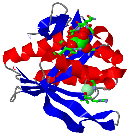
SCOP Domains (1, 1)
Asymmetric Unit
 |
CATH Domains (0, 0)| (no "CATH Domain" information available for 1S7F) |
Pfam Domains (0, 0)| (no "Pfam Domain" information available for 1S7F) |
Gene Ontology (5, 5)|
Asymmetric Unit(hide GO term definitions) Chain A (Q8ZPC0_SALTY | Q8ZPC0)
|
||||||||||||||||||||||||||||||||||||||||||||||||
Interactive Views
|
||||||||||||||||||||||||||||||||||||||||||||||||||||||||||||||||||||||||||||||||||||||||||||||||||||||||||||||||||||||||||||||||||||||||||||||||||||||
Still Images
|
||||||||||||||||
Databases
|
||||||||||||||||||||||||||||||||||||||||||||||||||||||||||||||||||||||||||||||||||||||||||||||||||||||||||||||||||||||||||||||||||||||||||||||||||||||||||||||||
Analysis Tools
|
|||||||||||||||||||||||||||||||||||||||||||||||||||||||||||||
Entries Sharing at Least One Protein Chain (UniProt ID)
Related Entries Specified in the PDB File
|
|