|
|
|
|
 Description
|
|
Compounds
|
||||||||||||||||||||||||||||||||||||||||||||||||||||
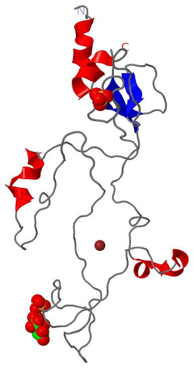
Chains, Units
Summary Information (see also Sequences/Alignments below) |
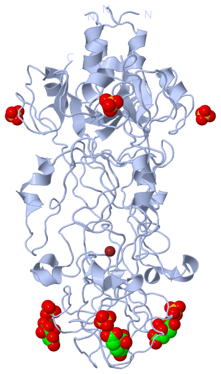
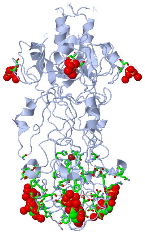
Ligands, Modified Residues, Ions (3, 4)| Asymmetric Unit (3, 4) Biological Unit 1 (2, 9) |
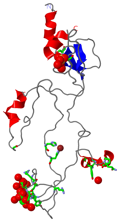
Sites (4, 4)
Asymmetric Unit (4, 4)
|
SS Bonds (0, 0)| (no "SS Bond" information available for 1OCY) |
Cis Peptide Bonds (0, 0)| (no "Cis Peptide Bond" information available for 1OCY) |
SAPs(SNPs)/Variants (0, 0)| (no "SAP(SNP)/Variant" information available for 1OCY) |
PROSITE Motifs (0, 0)| (no "PROSITE Motif" information available for 1OCY) |
Exons (0, 0)| (no "Exon" information available for 1OCY) |
Sequences/Alignments
Asymmetric UnitChain A from PDB Type:PROTEIN Length:198 aligned with FIB12_BPT4 | P10930 from UniProtKB/Swiss-Prot Length:527 Alignment length:198 339 349 359 369 379 389 399 409 419 429 439 449 459 469 479 489 499 509 519 FIB12_BPT4 330 RIVTQNEIDRTIPVGAIMMWAADSLPSDAWRFCHGGTVSASDCPLYASRIGTRYGGNPSNPGLPDMRGLFVRGSGRGSHLTNPNVNGNDQFGKPRLGVGCTGGYVGEVQIQQMSYHKHAGGFGEHDDLGAFGNTRRSNFVGTRKGLDWDNRSYFTNDGYEIDPESQRNSKYTLNRPELIGNETRPWNISLNYIIKVKE 527 SCOP domains d1ocya_ A: Receptor-binding domain of short tail fibre protein gp12 SCOP domains CATH domains 1ocyA01 A:330-396,A:519-527 1ocyA02 A:397-518 receptor-binding domain of the bacteriophage t4 short tail fibre, domain 2 1ocyA01 CATH domains Pfam domains ------------------------------------------------------------------------------------------------------------------------------------------------------------------------------------------------------ Pfam domains SAPs(SNPs) ------------------------------------------------------------------------------------------------------------------------------------------------------------------------------------------------------ SAPs(SNPs) PROSITE ------------------------------------------------------------------------------------------------------------------------------------------------------------------------------------------------------ PROSITE Transcript ------------------------------------------------------------------------------------------------------------------------------------------------------------------------------------------------------ Transcript 1ocy A 330 RVVTQNEIDRTIPVGAIMMWAADSLPSDAWRFCHGGTVSASDCPLYASRIGTRYGGSSSNPGLPDMRGLFVRGSGRGSHLTNPNVNGNDQFGKPRLGVGCTGGYVGEVQKQQMSYHKHAGGFGEYDDSGAFGNTRRSNFVGTRKGLDWDNRSYFTNDGYEIDPASQRNSRYTLNRPELIGNETRPWNISLNYIIKVKE 527 339 349 359 369 379 389 399 409 419 429 439 449 459 469 479 489 499 509 519 Chain A from PDB Type:PROTEIN Length:198 aligned with Q38160_BPT2 | Q38160 from UniProtKB/TrEMBL Length:527 Alignment length:198 339 349 359 369 379 389 399 409 419 429 439 449 459 469 479 489 499 509 519 Q38160_BPT2 330 RVVTQNEIDRTIPVGAIMMWAADSLPSDAWRFCHGGTVSASDCPLYASRIGTRYGGTSSNPGLPDMRGLFVRGSGRGSHLTNPNVNGNDQFGKPRLGVGCTGGYVGEVQKQQMSYHKHAGGFGEYDDSGAFGNTRRSNFVGTRKGLDWDNRSYFTNDGYEIDPASQRNSRYTLNRPELIGNETRPWNISLNYIIKVKE 527 SCOP domains d1ocya_ A: Receptor-binding domain of short tail fibre protein gp12 SCOP domains CATH domains 1ocyA01 A:330-396,A:519-527 1ocyA02 A:397-518 receptor-binding domain of the bacteriophage t4 short tail fibre, domain 2 1ocyA01 CATH domains Pfam domains ------------------------------------------------------------------------------------------------------------------------------------------------------------------------------------------------------ Pfam domains SAPs(SNPs) ------------------------------------------------------------------------------------------------------------------------------------------------------------------------------------------------------ SAPs(SNPs) PROSITE ------------------------------------------------------------------------------------------------------------------------------------------------------------------------------------------------------ PROSITE Transcript ------------------------------------------------------------------------------------------------------------------------------------------------------------------------------------------------------ Transcript 1ocy A 330 RVVTQNEIDRTIPVGAIMMWAADSLPSDAWRFCHGGTVSASDCPLYASRIGTRYGGSSSNPGLPDMRGLFVRGSGRGSHLTNPNVNGNDQFGKPRLGVGCTGGYVGEVQKQQMSYHKHAGGFGEYDDSGAFGNTRRSNFVGTRKGLDWDNRSYFTNDGYEIDPASQRNSRYTLNRPELIGNETRPWNISLNYIIKVKE 527 339 349 359 369 379 389 399 409 419 429 439 449 459 469 479 489 499 509 519
|
||||||||||||||||||||
SCOP Domains (1, 1)
Asymmetric Unit
 |
CATH Domains (2, 2)
Asymmetric Unit
|
Pfam Domains (0, 0)| (no "Pfam Domain" information available for 1OCY) |
Gene Ontology (5, 6)|
Asymmetric Unit(hide GO term definitions) Chain A (FIB12_BPT4 | P10930)
Chain A (Q38160_BPT2 | Q38160)
|
||||||||||||||||||||||||||||||||||||||||||||||||||||||||||||
Interactive Views
|
|||||||||||||||||||||||||||||||||||||||||||||||||||||||||||||||||||||||||||||||||||||||||||||||||||||||||||||||||||||||||||||||||||||||||||||||||||||||||||||||||||||||||||
Still Images
|
||||||||||||||||
Databases
|
||||||||||||||||||||||||||||||||||||||||||||||||||||||||||||||||||||||||||||||||||||||||||||||||||||||||||||||||||||||||||||||||||||||||||||||||||||||||||||||||||||||||||||||||||||||||||
Analysis Tools
|
||||||||||||||||||||||||||||||||||||||||||||||||||||||||||||||||||||||||
Entries Sharing at Least One Protein Chain (UniProt ID)
Related Entries Specified in the PDB File
|
|